


![Anti-HMGB1 antibody [EPR3506] 10µl](https://yunshiji.oss-cn-shenzhen.aliyuncs.com/202407/25/aeyqfwi4zsl.jpg)
![Anti-HMGB1 antibody [EPR3506] 100µl](https://yunshiji.oss-cn-shenzhen.aliyuncs.com/202407/25/skpaxrflv4o.jpg)
![Anti-HMGA1 antibody [EPR7839] 10µl](https://yunshiji.oss-cn-shenzhen.aliyuncs.com/202407/25/d00mjfdocky.jpg)
![Anti-HMGA1 antibody [EPR7839] 40µl](https://yunshiji.oss-cn-shenzhen.aliyuncs.com/202407/25/tgearga4etp.jpg)
![Anti-HMGA1 antibody [EPR7839] 100µl](https://yunshiji.oss-cn-shenzhen.aliyuncs.com/202407/25/epbjz5juk2g.jpg)
![Anti-HMGA1 antibody [EPR16649] 10µl](https://yunshiji.oss-cn-shenzhen.aliyuncs.com/202407/25/xcemuxdxk51.jpg)
详细说明
概述
产品名称Anti-NAK/TBK1抗体[EPR2867(2)-19]
描述
兔单克隆抗体[EPR2867(2)-19] to NAK/TBK1
经测试应用WB,IHC-P,Flow Cyt
种属反应性
与反应: Mouse, Rat, Human
免疫原
Synthetic peptide (the amino acid sequence is considered to be commercially sensitive) corresponding to Human NAK/TBK1 aa 150-250.
阳性对照
HeLa, U937, HepG2, and 293T cell lysates, Human testis tissue, HeLa cells
常规说明
This product is a recombinant rabbit monoclonal antibody.
We are constantly working hard to ensure we provide our customers with best in class antibodies. As a result of this work we are pleased to now offer this antibody in purified format. We are in the process of updating our datasheets. The purified format is designated ‘PUR’ on our product labels. If you have any questions regarding this update, please contact our Scientific Support team.
Produced using Abcam’s RabMAb® technology. RabMAb® technology is covered by the following U.S. Patents, No. 5,675,063 and/or 7,429,487.
性能
形式Liquid
存放说明Shipped at 4°C. Store at -20°C. Stable for 12 months at -20°C.
存储溶液pH: 7.20
Preservative: 0.01% Sodium azide
Constituents: 59% PBS, 40% Glycerol, 0.05% BSA纯度Protein A purified
克隆单克隆
克隆编号EPR2867(2)-19
同种型IgG
研究领域
Epigenetics and Nuclear Signaling
Nuclear Signaling Pathways
NFkB pathway
Signal Transduction
Signaling Pathway
Nuclear Signaling
NFkB Pathway
<a javascript:void(0)>Signal Transduction
Protein Phosphorylation
Ser / Thr Kinases
Other Kinases
Anti-NAK/TBK1 antibody [EPR2867(2)-19] 图像
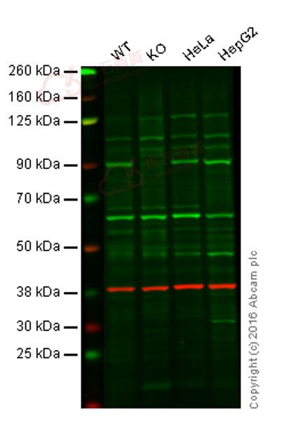
![Western blot - Anti-NAK/TBK1 antibody [EPR2867(2)-19] (ab109735)](http://img.lianshimall.com/statics/attachment/goods/pl20160426/abcamMainImgPrimary/detail/ab10/ab109735OWB.jpg)
Western blot - Anti-NAK/TBK1 antibody [EPR2867(2)-19] (ab109735)
Predicted band size : 84 kDa
Lane 1: Wild-type HAP1 cell lysate (20 µg)
Lane 2: NAK/TBK1 knockout HAP1 cell lysate (20 µg)
Lane 3: HeLa cell lysate (20 µg)
Lane 4: HepG2 cell lysate (20 µg)
Lanes 1 - 4: Merged signal (red and green). Green - ab109735 observed at 90 kDa. Red - loading control, ab8245, observed at 37 kDa.
ab109735 was shown to recognize NAK/TBK1 when NAK/TBK1 knockout samples were used, along with additional cross-reactive bands. Wild-type and NAK/TBK1 knockout samples were subjected to SDS-PAGE. ab109735 and ab8245 (loading control to GAPDH) were diluted at 1/1000 and 1/10 000 respectively and incubated overnight at 4°C. Blots were developed with goat anti-rabbit IgG (H + L) and goat anti-mouse IgG (H + L) secondary antibodies at 1/10 000 dilution for 1 h at room temperature before imaging.![Western blot - Anti-NAK/TBK1 antibody [EPR2867(2)-19] (ab109735)](http://img.lianshimall.com/statics/attachment/goods/pl20160426/abcamMainImgPrimary/detail/ab10/ab109735-WB.jpg)
Western blot - Anti-NAK/TBK1 antibody [EPR2867(2)-19] (ab109735)
Anti-NAK/TBK1 antibody [EPR2867(2)-19] (ab109735) at 1/4000 dilution + HeLa cell lysate at 10 µg
Secondary
Goat Anti-Rabbit IgG, (H+L), HRP-conjugated at 1/1000 dilution
Predicted band size : 84 kDa
Blocking buffer and concentration: 5% NFDM/TBST.
Diluting buffer and concentration: 5% NFDM /TBST.
![Immunohistochemistry (Formalin/PFA-fixed paraffin-embedded sections) - Anti-NAK/TBK1 antibody [EPR2867(2)-19] (ab109735)](http://img.lianshimall.com/statics/attachment/goods/pl20160426/abcamMainImgPrimary/detail/ab10/ab109735IHC.jpg)
Immunohistochemistry (Formalin/PFA-fixed paraffin-embedded sections) - Anti-NAK/TBK1 antibody [EPR2867(2)-19] (ab109735)
Immunohistochemical analysis of paraffin-embedded human testis tissue using ab109735 at a dilution of 1/250.
Immunohistochemistry (Formalin/PFA-fixed paraffin-embedded sections) - Anti-NAK/TBK1 antibody [EPR2867(2)-19] (ab109735)
ab109735 staining NAK/TBK1 in Human liver tissue sections by Immunohistochemistry (IHC-P - paraformaldehyde-fixed, paraffin-embedded sections). Tissue was fixed and paraffin-embedded, antigen retrieval was by heat mediation in Tris/EDTA buffer pH9. Samples were incubated with primary antibody (1/800). An undiluted HRP-conjugated anti-rabbit IgG was used as the secondary antibody. Tissue counterstained with Hematoxylin.
![Flow Cytometry - Anti-NAK/TBK1 antibody [EPR2867(2)-19] (ab109735)](http://img.lianshimall.com/statics/attachment/goods/pl20160426/abcamMainImgPrimary/detail/ab10/ab109735low.jpg)
Flow Cytometry - Anti-NAK/TBK1 antibody [EPR2867(2)-19] (ab109735)
Overlay histogram showing HeLa cells stained with ab109735 (red line) at 1/90 dilution. The cells were fixed with 2% paraformaldehyde. The secondary antibody used was a FITC conjugated goat anti-rabbit IgG at 1/150 dilution. Isotype control antibody (green line) was rabbit monoclonal IgG used under the same conditions.
![Flow Cytometry - Anti-NAK/TBK1 antibody [EPR2867(2)-19] (ab109735)](http://img.lianshimall.com/statics/attachment/goods/pl20160426/abcamMainImgPrimary/detail/ab10/ab1097355FC.jpg)
Flow Cytometry - Anti-NAK/TBK1 antibody [EPR2867(2)-19] (ab109735)
Overlay histogram showing HeLa cells stained with ab109735 (red line). The cells were fixed with 80% methanol (5 min) and then permeabilized with 0.1% PBS-Tween for 20 min. The cells were then incubated in 1x PBS / 10% normal goat serum / 0.3M glycine to block non-specific protein-protein interactions followed by the antibody (ab109735, 1/1000 dilution) for 30 min at 22°C. The secondary antibody used was Alexa Fluor® 488 goat anti-rabbit IgG (H&L) (ab150077) at 1/2000 dilution for 30 min at 22°C. Isotype control antibody (black line) was rabbit IgG (monoclonal) (0.1μg/1x106 cells) used under the same conditions. Unlabelled sample (blue line) was also used as a control. Acquisition of >5,000 events were collected using a 20mW Argon ion laser (488nm) and 525/30 bandpass filter.
![Western blot - Anti-NAK/TBK1 antibody [EPR2867(2)-19] (ab109735)](http://img.lianshimall.com/statics/attachment/goods/pl20160426/abcamMainImgPrimary/detail/ab10/ab1097355-1.jpg)
Western blot - Anti-NAK/TBK1 antibody [EPR2867(2)-19] (ab109735)
All lanes : Anti-NAK/TBK1 antibody [EPR2867(2)-19] (ab109735) at 1/1000 dilution
Lane 1 : HeLa cell lysate
Lane 2 : U937 cell lysate
Lane 3 : HepG2 cell lysate
Lane 4 : 293T cell lysate
Lysates/proteins at 10 µg per lane.
Predicted band size : 84 kDa


 粤公网安备44196802000105号