


![Anti-JNK1 antibody [EPR140(2)] 10µl](https://yunshiji.oss-cn-shenzhen.aliyuncs.com/202407/25/iyztyimz0ge.jpg)
![Anti-JNK1 antibody [EPR140(2)] 40µl](https://yunshiji.oss-cn-shenzhen.aliyuncs.com/202407/25/53qxai2no5j.jpg)
![Anti-JNK1 antibody [EPR140(2)] 100µl](https://yunshiji.oss-cn-shenzhen.aliyuncs.com/202407/25/bi1jw5v3pkp.jpg)
![Anti-JNK1 + JNK3 antibody [EPR16797-194] 10µl](https://yunshiji.oss-cn-shenzhen.aliyuncs.com/202407/25/s00dbqjm5f4.jpg)
![Anti-JNK1 + JNK3 antibody [EPR16797-194] 40µl](https://yunshiji.oss-cn-shenzhen.aliyuncs.com/202407/25/5tqlusihwkd.jpg)
![Anti-JNK1 + JNK3 antibody [EPR16797-194] 100µl](https://yunshiji.oss-cn-shenzhen.aliyuncs.com/202407/25/stuzawgabrf.jpg)
详细说明
概述
产品名称Anti-MTHFR抗体[EPR19781]
描述
兔单克隆抗体[EPR19781] to MTHFR
经测试应用IP,ICC/IF,WB
种属反应性
与反应: Mouse, Human
免疫原
Recombinant fragment within Human MTHFR aa 1-300. The exact sequence is proprietary.
Database link: P42898Run BLAST with
 Run BLAST with
阳性对照
WB: Human fetal brain, fetal heart, fetal kidney, spleen and fetal liver lysates; Jurkat, HT-29, HEK-293, RAW 264.7 and NIH/3T3 whole cell lysates; Mouse placenta and kidney lysates. ICC/IF: Jurkat and RAW 264.7 cells. IP: HT-29 whole cell lysate.
常规说明
This product is a recombinant rabbit monoclonal antibody.
Produced using Abcam's RabMAb® technology. RabMAb® technology is covered by the following U.S. Patents, No. 5, 675, 063 and/or 7, 429, 487.
性能
形式Liquid
存放说明Shipped at 4°C. Store at +4°C short term (1-2 weeks). Upon delivery aliquot. Store at -20°C long term. Avoid freeze / thaw cycle.
存储溶液Preservative: 0.01% Sodium azide
Constituents: 59% PBS, 40% Glycerol, 0.05% BSA纯度Protein A purified
克隆单克隆
克隆编号EPR19781
同种型IgG
研究领域
Metabolism
Pathways and Processes
Metabolic signaling pathways
Amino acid metabolism
Signal Transduction
Metabolism
Amino Acids
Anti-MTHFR antibody [EPR19781] 图像
![Western blot - Anti-MTHFR antibody [EPR19781] (ab203786)](http://img.lianshimall.com/statics/attachment/goods/pl20160426/abcamMainImgPrimary/detail/ab2/ab203786WBa.jpg)
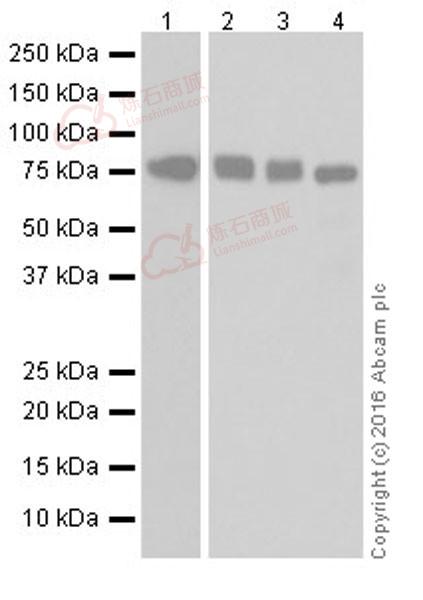
Western blot - Anti-MTHFR antibody [EPR19781] (ab203786)
All lanes : Anti-MTHFR antibody [EPR19781] (ab203786) at 1/1000 dilution
Lane 1 : Human fetal brain lysate
Lane 2 : Human fetal heart lysate
Lane 3 : Human fetal kidney lysate
Lane 4 : Human spleen lysate
Lysates/proteins at 10 µg per lane.
Secondary
Goat Anti-Rabbit IgG Peroxidase Conjugate, specific to the non-reduced form of IgG at 1/10000 dilution
Predicted band size : 75 kDa
Observed band size : 75 kDa
Blocking/Dilution buffer: 5% NFDM/TBST.
Exposure time: Lane 1: 3 minutes; Lane 2,3 and 4: 30 seconds.
![Western blot - Anti-MTHFR antibody [EPR19781] (ab203786)](http://img.lianshimall.com/statics/attachment/goods/pl20160426/abcamMainImgPrimary/detail/ab2/ab203786WBb.jpg)
Western blot - Anti-MTHFR antibody [EPR19781] (ab203786)
Lanes 1 - 3 : Anti-MTHFR antibody [EPR19781] (ab203786) at 1/1000 dilution
Lanes 4 - 5 : Anti-MTHFR antibody [EPR19781] (ab203786) at 1/5000 dilution
Lane 1 : Jurkat (Human T cell leukemia cell line from peripheral blood) whole cell lysate
Lane 2 : HT-29 (Human colorectal adenocarcinoma cell line) whole cell lysate
Lane 3 : Human fetal liver lysate
Lane 4 : HEK-293 (Human epithelial cell line from embryonic kidney) whole cell lysate
Lane 5 : Mouse placenta lysate
Lysates/proteins at 20 µg per lane.
Secondary
Goat Anti-Rabbit IgG H&L (HRP) (ab97051) at 1/100000 dilution
Predicted band size : 75 kDa
Observed band size : 75 kDa
Exposure time : 3 minutesBlocking/Dilution buffer: 5% NFDM/TBST.
![Western blot - Anti-MTHFR antibody [EPR19781] (ab203786)](http://img.lianshimall.com/statics/attachment/goods/pl20160426/abcamMainImgPrimary/detail/ab2/ab203786WBc.jpg)
Western blot - Anti-MTHFR antibody [EPR19781] (ab203786)
All lanes : Anti-MTHFR antibody [EPR19781] (ab203786) at 1/1000 dilution
Lane 1 : RAW 264.7 (Mouse macrophage cell line transformed with Abelson murine leukemia virus) whole cell lysate
Lane 2 : NIH/3T3 (Mouse embryonic fibroblast cell line) whole cell lysate
Lane 3 : Mouse kidney lysate
Lysates/proteins at 20 µg per lane.
Secondary
Goat Anti-Rabbit IgG H&L (HRP) (ab97051) at 1/100000 dilution
Predicted band size : 75 kDa
Observed band size : 75 kDa
Exposure time : 3 minutesBlocking/Dilution buffer: 5% NFDM/TBST.
![Immunocytochemistry/ Immunofluorescence - Anti-MTHFR antibody [EPR19781] (ab203786)](http://img.lianshimall.com/statics/attachment/goods/pl20160426/abcamMainImgPrimary/detail/ab2/ab203786IFa.jpg)
Immunocytochemistry/ Immunofluorescence - Anti-MTHFR antibody [EPR19781] (ab203786)
Immunofluorescent analysis of 4% paraformaldehyde-fixed, 0.1% Triton X-100 permeabilized Jurkat (Human T cell leukemia cell line from peripheral blood) cells labeling MTHFR with ab203786 at 1/100 dilution, followed by Goat Anti-Rabbit IgG (Alexa Fluor® 488) (ab150077) secondary antibody at 1/1000 dilution (green).
Confocal image showing cytoplasmic staining on Jurkat cell line.
The nuclear counterstain is DAPI (blue).
Tubulin is detected with Anti-alpha Tubulin antibody [DM1A] - Loading Control (ab7291) at 1/1000 dilution and Goat Anti-Mouse IgG H&L (Alexa Fluor® 594) preadsorbed (ab150120) at 1/1000 dilution (red).
The negative controls are as follows:-
-ve control 1: ab203786 at 1/100 dilution followed by ab150120 at 1/1000 dilution.
-ve control 2: ab7291 at 1/1000 dilution followed by ab150077 at 1/1000 dilution.
![Immunocytochemistry/ Immunofluorescence - Anti-MTHFR antibody [EPR19781] (ab203786)](http://img.lianshimall.com/statics/attachment/goods/pl20160426/abcamMainImgPrimary/detail/ab2/ab203786IFb.jpg)
Immunocytochemistry/ Immunofluorescence - Anti-MTHFR antibody [EPR19781] (ab203786)
Immunofluorescent analysis of 4% paraformaldehyde-fixed, 0.1% Triton X-100 permeabilized RAW 264.7 (Mouse macrophage cell line transformed with Abelson murine leukemia virus) cells labeling MTHFR with ab203786 at 1/100 dilution, followed by Goat Anti-Rabbit IgG (Alexa Fluor® 488) (ab150077) secondary antibody at 1/1000 dilution (green).
Confocal image showing cytoplasmic staining on RAW 264.7 cell line.
The nuclear counterstain is DAPI (blue).
Tubulin is detected with Anti-alpha Tubulin antibody [DM1A] - Loading Control (ab7291) at 1/1000 dilution and Goat Anti-Mouse IgG H&L (Alexa Fluor® 594) preadsorbed (ab150120) at 1/1000 dilution (red).
The negative controls are as follows:-
-ve control 1: ab203786 at 1/100 dilution followed by ab150120 at 1/1000 dilution.
-ve control 2: ab7291 at 1/1000 dilution followed by ab150077 at 1/1000 dilution.
![Immunoprecipitation - Anti-MTHFR antibody [EPR19781] (ab203786)](http://img.lianshimall.com/statics/attachment/goods/pl20160426/abcamMainImgPrimary/detail/ab2/ab2037866IP.jpg)
Immunoprecipitation - Anti-MTHFR antibody [EPR19781] (ab203786)
MTHFR was immunoprecipitated from 0.35mg of HT-29 (Human colorectal adenocarcinoma cell line) whole cell lysate with ab203786 at 1/30 dilution.
Western blot was performed from the immunoprecipitate using ab203786 at 1/500 dilution.
VeriBlot for IP secondary antibody (HRP) (ab131366), was used as secondary antibody at 1/1000 dilution.
Lane 1: HT-29 whole cell lysate, 10µg (Input).
Lane 2: ab203786 IP in HT-29 whole cell lysate.
Lane 3: Rabbit IgG, monoclonal [EPR25A] - Isotype Control (ab172730) instead of ab203786 in HT-29 whole cell lysate.
Blocking and dilution buffer and concentration: 5% NFDM/TBST.
Exposure time: 10 seconds.


 粤公网安备44196802000105号