


![Anti-Histone H3 (tri methyl K79) antibody [EPR17468(2)] 10µl](https://yunshiji.oss-cn-shenzhen.aliyuncs.com/202407/25/olglrpt5cyw.jpg)
![Anti-MUC4 antibody [EPR9308] 10µl](https://yunshiji.oss-cn-shenzhen.aliyuncs.com/202407/25/j0b0sfrccss.jpg)
![Anti-MUC4 antibody [EPR9308] 40µl](https://yunshiji.oss-cn-shenzhen.aliyuncs.com/202407/25/erj2134plp2.jpg)
![Anti-MUC4 antibody [EPR9308] 100µl](https://yunshiji.oss-cn-shenzhen.aliyuncs.com/202407/25/lyimit0rrvb.jpg)
![Anti-MUC4 antibody [EPR16237] 10µl](https://yunshiji.oss-cn-shenzhen.aliyuncs.com/202407/25/l1vsfnefyix.jpg)
![Anti-MUC4 antibody [EPR16237] 40µl](https://yunshiji.oss-cn-shenzhen.aliyuncs.com/202407/25/hylgpiq50zi.jpg)
详细说明
概述
产品名称Anti-Histone H3抗体[EPR17785]
描述
兔单克隆抗体[EPR17785] to Histone H3
经测试应用WB,ICC/IF,Flow Cyt,IHC-P
种属反应性
与反应: Mouse, Rat, Human
免疫原
Synthetic peptide (the amino acid sequence is considered to be commercially sensitive) within Human Histone H3 aa 1-100. The exact sequence is proprietary.
Database link: P68431阳性对照
WB: HeLa, HEK293, A375 and NIH/3T3 cell lysates; Human fetal brain, mouse kidney and rat brain lysates. IHC-P: Human cervix carcinoma, Human kidney, mouse colon and rat stomach tissues. ICC/IF: HeLa cells. Flow Cyt: HeLa cells.
常规说明
This product is a recombinant rabbit monoclonal antibody.
Produced using Abcam’s RabMAb® technology. RabMAb® technology is covered by the following U.S. Patents, No. 5,675,063 and/or 7,429,487.
性能
形式Liquid
存放说明Shipped at 4°C. Store at +4°C short term (1-2 weeks). Upon delivery aliquot. Store at -20°C long term. Avoid freeze / thaw cycle.
存储溶液Preservative: 0.01% Sodium azide
Constituents: 59% PBS, 40% Glycerol, 0.05% BSA纯度Protein A purified
克隆单克隆
克隆编号EPR17785
同种型IgG
研究领域
Epigenetics and Nuclear Signaling
Histones
H3
Unmodified
Anti-Histone H3 antibody [EPR17785] 图像
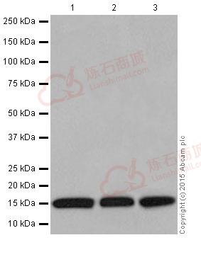
![Western blot - Anti-Histone H3 antibody [EPR17785] (ab201456)](http://img.lianshimall.com/statics/attachment/goods/pl20160426/abcamMainImgPrimary/detail/ab2/ab201456WBa.jpg)
Western blot - Anti-Histone H3 antibody [EPR17785] (ab201456)
All lanes : Anti-Histone H3 antibody [EPR17785] (ab201456) at 1/2000 dilution
Lane 1 : HeLa (Human epithelial cells from cervix adenocarcinoma) cell lysate
Lane 2 : HEK293 (Human embryonic kidney) cell lysate
Lane 3 : A375 (Human malignant melanoma) cell lysate
Lysates/proteins at 20 µg per lane.
Secondary
Goat Anti-Rabbit IgG, (H+L), Peroxidase conjugated at 1/1000 dilution
Predicted band size : 15 kDa
Observed band size : 15 kDa
Exposure time : 15 secondsBlocking/Dilution buffer: 5% NFDM/TBST.
![Western blot - Anti-Histone H3 antibody [EPR17785] (ab201456)](http://img.lianshimall.com/statics/attachment/goods/pl20160426/abcamMainImgPrimary/detail/ab2/ab201456WBb.jpg)
Western blot - Anti-Histone H3 antibody [EPR17785] (ab201456)
Anti-Histone H3 antibody [EPR17785] (ab201456) at 1/2000 dilution + Human fetal brain lysate at 10 µg
Secondary
Anti-Rabbit IgG (HRP), specific to the non-reduced form of IgG at 1/1000 dilution
Predicted band size : 15 kDa
Observed band size : 15 kDa
Exposure time : 3 minutesBlocking/Dilution buffer: 5% NFDM/TBST.
![Western blot - Anti-Histone H3 antibody [EPR17785] (ab201456)](http://img.lianshimall.com/statics/attachment/goods/pl20160426/abcamMainImgPrimary/detail/ab2/ab201456WBc.jpg)
Western blot - Anti-Histone H3 antibody [EPR17785] (ab201456)
All lanes : Anti-Histone H3 antibody [EPR17785] (ab201456) at 1/2000 dilution
Lane 1 : Mouse kidney lysate
Lane 2 : Rat brain lysate
Lane 3 : NIH/3T3 (Mouse embyro fibroblast cells) cell lysate
Lysates/proteins at 10 µg per lane.
Secondary
Goat Anti-Rabbit IgG, (H+L), Peroxidase conjugated at 1/1000 dilution
Predicted band size : 15 kDa
Observed band size : 15 kDa
Exposure time : 1 minuteBlocking/Dilution buffer: 5% NFDM/TBST.
![Immunohistochemistry (Formalin/PFA-fixed paraffin-embedded sections) - Anti-Histone H3 antibody [EPR17785] (ab201456)](http://img.lianshimall.com/statics/attachment/goods/pl20160426/abcamMainImgPrimary/detail/ab2/ab201456HCa.jpg)
Immunohistochemistry (Formalin/PFA-fixed paraffin-embedded sections) - Anti-Histone H3 antibody [EPR17785] (ab201456)
Immunohistochemical analysis of paraffin-embedded Human cervix carcinoma tissue labeling Histone H3 with ab201456 at 1/200 dilution, followed by Goat Anti-Rabbit IgG H&L (HRP) (ab97051) secondary antibody at 1/500 dilution.
Nuclear staining on Human cervix carcinoma tissue is observed.
Counter stained with Hematoxylin.
Secondary antibody only control: Used PBS instead of primary antibody, secondary antibody is Goat Anti-Rabbit IgG H&L (HRP) (ab97051) at 1/500 dilution.
![Immunohistochemistry (Formalin/PFA-fixed paraffin-embedded sections) - Anti-Histone H3 antibody [EPR17785] (ab201456)](http://img.lianshimall.com/statics/attachment/goods/pl20160426/abcamMainImgPrimary/detail/ab2/ab201456HCb.jpg)
Immunohistochemistry (Formalin/PFA-fixed paraffin-embedded sections) - Anti-Histone H3 antibody [EPR17785] (ab201456)
Immunohistochemical analysis of paraffin-embedded Human kidney tissue labeling Histone H3 with ab201456 at 1/200 dilution, followed by Goat Anti-Rabbit IgG H&L (HRP) (ab97051) secondary antibody at 1/500 dilution.
Nuclear staining on Human kidney tissue is observed.
Counter stained with Hematoxylin.
Secondary antibody only control: Used PBS instead of primary antibody, secondary antibody is Goat Anti-Rabbit IgG H&L (HRP) (ab97051) at 1/500 dilution.
![Immunohistochemistry (Formalin/PFA-fixed paraffin-embedded sections) - Anti-Histone H3 antibody [EPR17785] (ab201456)](http://img.lianshimall.com/statics/attachment/goods/pl20160426/abcamMainImgPrimary/detail/ab2/ab201456HCc.jpg)
Immunohistochemistry (Formalin/PFA-fixed paraffin-embedded sections) - Anti-Histone H3 antibody [EPR17785] (ab201456)
Immunohistochemical analysis of paraffin-embedded Mouse colon tissue labeling Histone H3 with ab201456 at 1/200 dilution, followed by Goat Anti-Rabbit IgG H&L (HRP) (ab97051) secondary antibody at 1/500 dilution.
Nuclear staining on mouse colon tissue is observed.
Counter stained with Hematoxylin.
Secondary antibody only control: Used PBS instead of primary antibody, secondary antibody is Goat Anti-Rabbit IgG H&L (HRP) (ab97051) at 1/500 dilution.
![Immunohistochemistry (Formalin/PFA-fixed paraffin-embedded sections) - Anti-Histone H3 antibody [EPR17785] (ab201456)](http://img.lianshimall.com/statics/attachment/goods/pl20160426/abcamMainImgPrimary/detail/ab2/ab201456HCd.jpg)
Immunohistochemistry (Formalin/PFA-fixed paraffin-embedded sections) - Anti-Histone H3 antibody [EPR17785] (ab201456)
Immunohistochemical analysis of paraffin-embedded Rat stomach tissue labeling Histone H3 with ab201456 at 1/200 dilution, followed by Goat Anti-Rabbit IgG H&L (HRP) (ab97051) secondary antibody at 1/500 dilution.
Nuclear staining on rat stomach tissue is observed.
Counter stained with Hematoxylin.
Secondary antibody only control: Used PBS instead of primary antibody, secondary antibody is Goat Anti-Rabbit IgG H&L (HRP) (ab97051) at 1/500 dilution.
![Immunocytochemistry/ Immunofluorescence - Anti-Histone H3 antibody [EPR17785] (ab201456)](http://img.lianshimall.com/statics/attachment/goods/pl20160426/abcamMainImgPrimary/detail/ab2/ab2014566IF.jpg)
Immunocytochemistry/ Immunofluorescence - Anti-Histone H3 antibody [EPR17785] (ab201456)
Immunofluorescent analysis of 4% paraformaldehyde-fixed, 0.1% Triton-X100 permeabilized HeLa (Human epithelial cells from cervix adenocarcinoma) cells labeling Histone H3 with ab201456 at 1/800 dilution, followed by AlexaFluor®488 Goat anti-Rabbit secondary antibody (ab150077) at 1/500 dilution (green).
Confocal image showing nuclear staining on HeLa cell line.
The nuclear counter stain is DAPI (blue).
Tubulin is stained with ab7291 anti-Tubulin (mouse mAb) at 1/1000 dilution, followed by AlexaFluor®594 Goat anti-Mouse secondary antibody (ab150120) at 1/500 dilution (red).
-ve control 1: ab201456 at 1/800 dilution followed by AlexaFluor®594 Goat anti-Mouse secondary antibody (ab150120) at 1/500 dilution.
-ve control 2: ab7291 anti-Tubulin (mouse mAb) at 1/1000 dilution, followed by AlexaFluor®488 Goat anti-Rabbit secondary antibody (ab150077) at 1/500 dilution.![Flow Cytometry - Anti-Histone H3 antibody [EPR17785] (ab201456)](http://img.lianshimall.com/statics/attachment/goods/pl20160426/abcamMainImgPrimary/detail/ab2/ab2014566FC.jpg)
Flow Cytometry - Anti-Histone H3 antibody [EPR17785] (ab201456)
Flow cytometric analysis of 2% paraformaldehyde-fixed HeLa (Human epithelial cells from cervix adenocarcinoma) cells labeling Histone H3 with ab201456 at 1/100 dilution (red) compared with a rabbit monoclonal IgG isotype control (black) and an unlabelled control (cells without incubation with primary antibody and secondary antibody; blue). Goat anti rabbit IgG (FITC) at 1/150 dilution was used as the secondary antibody.


 粤公网安备44196802000105号