


![Anti-Histone H3 (tri methyl K79) antibody [EPR17468(2)] 10µl](https://yunshiji.oss-cn-shenzhen.aliyuncs.com/202407/25/olglrpt5cyw.jpg)
![Anti-MUC4 antibody [EPR9308] 10µl](https://yunshiji.oss-cn-shenzhen.aliyuncs.com/202407/25/j0b0sfrccss.jpg)
![Anti-MUC4 antibody [EPR9308] 40µl](https://yunshiji.oss-cn-shenzhen.aliyuncs.com/202407/25/erj2134plp2.jpg)
![Anti-MUC4 antibody [EPR9308] 100µl](https://yunshiji.oss-cn-shenzhen.aliyuncs.com/202407/25/lyimit0rrvb.jpg)
![Anti-MUC4 antibody [EPR16237] 10µl](https://yunshiji.oss-cn-shenzhen.aliyuncs.com/202407/25/l1vsfnefyix.jpg)
![Anti-MUC4 antibody [EPR16237] 40µl](https://yunshiji.oss-cn-shenzhen.aliyuncs.com/202407/25/hylgpiq50zi.jpg)
详细说明
概述
产品名称Anti-Histone H3.3抗体[EPR17899] - ChIP Grade
描述
兔单克隆抗体[EPR17899] to Histone H3.3 - ChIP Grade
特异性The immunogen sequence used for this antibody is 100% identical to the protein sequence shown for H3.X and H3.Y in Szenker et al., 2011 (PubMed ID 21263457). These H3.X and H3.Y protein sequences have not been reviewed by UniProt. For further information on this antibody, please contact our technical support team.
经测试应用ICC/IF,ChIP,IHC-P,WB,Dot Blot
种属反应性
与反应: Mouse, Rat, Human
免疫原
Synthetic peptide (the amino acid sequence is considered to be commercially sensitive) within Human Histone H3.3 aa 50 to the C-terminus. The exact sequence is proprietary.
Database link: P84243阳性对照
WB: HeLa and NIH/3T3 whole cell lysates. IHC-P: Human colon, Mouse stomach and Rat colon tissues. ICC/IF: HeLa cells. ChIP: Chromatin prepared from HeLa cells.
常规说明
This product is a recombinant rabbit monoclonal antibody.
Produced using Abcam’s RabMAb® technology. RabMAb® technology is covered by the following U.S. Patents, No. 5,675,063 and/or 7,429,487.
性能
形式Liquid
存放说明Shipped at 4°C. Store at +4°C short term (1-2 weeks). Upon delivery aliquot. Store at -20°C long term. Avoid freeze / thaw cycle.
存储溶液Preservative: 0.01% Sodium azide
Constituents: 59% PBS, 0.05% BSA, 40% Glycerol纯度Protein A purified
克隆单克隆
克隆编号EPR17899
同种型IgG
研究领域
Epigenetics and Nuclear Signaling
Histones
Variants
Anti-Histone H3.3 antibody [EPR17899] - ChIP Grade 图像
![Western blot - Anti-Histone H3.3 antibody [EPR17899] (ab176840)](http://img.lianshimall.com/statics/attachment/goods/pl20160426/abcamMainImgPrimary/detail/ab17/ab176840899.jpg)
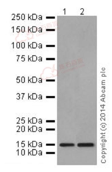
Western blot - Anti-Histone H3.3 antibody [EPR17899] (ab176840)
All lanes : Anti-Histone H3.3 antibody [EPR17899] - ChIP Grade (ab176840) at 1/1000 dilution
Lane 1 : HeLa (Human epithelial cells from cervix adenocarcinoma) whole cell lysate
Lane 2 : NIH/3T3 (Mouse embyro fibroblast cells) whole cell lysate
Lysates/proteins at 10 µg per lane.
Secondary
Goat Anti-Rabbit IgG, (H+L), Peroxidase conjugated at 1/1000 dilution
Predicted band size : 15 kDa
Observed band size : 15 kDa
Exposure time : 3 minutesBlocking/Dilution buffer: 5% NFDM/TBST.
![Dot Blot - Anti-Histone H3.3 antibody [EPR17899] (ab176840)](http://img.lianshimall.com/statics/attachment/goods/pl20160426/abcamMainImgPrimary/detail/ab17/ab1768409DB.jpg)
Dot Blot - Anti-Histone H3.3 antibody [EPR17899] (ab176840)
Dot blot analysis of Histone H3.3 peptide (Lane 1), and Histone H3.1 peptide (Lane 2), labeled using ab176840 at 1/1000 dilution, followed by Goat Anti-Rabbit IgG, (H+L), Peroxidase conjugated secondary antibody at 1/1000 dilution.
Blocking/Dilution buffer: 5% NFDM/TBST.
![Western blot - Anti-Histone H3.3 antibody [EPR17899] (ab176840)](http://img.lianshimall.com/statics/attachment/goods/pl20160426/abcamMainImgPrimary/detail/ab17/ab176840WBb.jpg)
Western blot - Anti-Histone H3.3 antibody [EPR17899] (ab176840)
All lanes : Anti-Histone H3.3 antibody [EPR17899] - ChIP Grade (ab176840) at 1/1000 dilution
Lane 1 : HeLa (Human epithelial cells from cervix adenocarcinoma) whole cell lysate
Lane 2 : HeLa (Human epithelial cells from cervix adenocarcinoma) whole cell lysate with Histone H3.3 peptide
Lane 3 : HeLa (Human epithelial cells from cervix adenocarcinoma) whole cell lysate with Histone H3.1 peptide
Lane 4 : NIH/3T3 (Mouse embyro fibroblast cells) whole cell lysate
Lane 5 : NIH/3T3 (Mouse embyro fibroblast cells) whole cell lysate with Histone H3.3 peptide
Lane 6 : NIH/3T3 (Mouse embyro fibroblast cells) whole cell lysate with Histone H3.1 peptide
Lysates/proteins at 10 µg per lane.
Secondary
Goat Anti-Rabbit IgG, (H+L), Peroxidase conjugated at 1/1000 dilution
Predicted band size : 15 kDa
Observed band size : 15 kDa
Exposure time : 3 minutesBlocking experiment by pre-incubating Histone H3.3 peptide with the antibody in lanes 2 and 5 and Histone H3.1 peptide in lanes 3 and 6 to show the antibody specificity against Histone H3.3.
Blocking/Dilution buffer: 5% NFDM/TBST.
![ChIP - Anti-Histone H3.3 antibody [EPR17899] (ab176840)](http://img.lianshimall.com/statics/attachment/goods/pl20160426/abcamMainImgPrimary/detail/ab17/ab176840hIP.jpg)
ChIP - Anti-Histone H3.3 antibody [EPR17899] (ab176840)
Chromatin was prepared from HeLa (Human epithelial cells from cervix adenocarcinoma) cells according to the Abcam X-ChIP protocol. Cells were fixed with formaldehyde for 10 minutes. The ChIP was performed with 25µg of chromatin, 2µg of ab176840 (blue), and 20µl of Anti rabbit IgG sepharose beads. 2μg of rabbit normal IgG was added to the beads control (yellow). The immunoprecipitated DNA was quantified by real time PCR (Sybr green approach).
![Immunocytochemistry/ Immunofluorescence - Anti-Histone H3.3 antibody [EPR17899] (ab176840)](http://img.lianshimall.com/statics/attachment/goods/pl20160426/abcamMainImgPrimary/detail/ab17/ab176840ged.jpg)
Immunocytochemistry/ Immunofluorescence - Anti-Histone H3.3 antibody [EPR17899] (ab176840)
Immunofluorescent analysis of 4% paraformaldehyde-fixed, 0.1% Triton X-100 permeabilized HeLa (Human epithelial cells from cervix adenocarcinoma) cells labeling Histone H3.3 with ab176840 at 1/1000 dilution, followed by Goat anti-rabbit IgG (Alexa Fluor® 488) (ab150077) secondary antibody at 1/500 dilution (green). Confocal image showing nuclear staining on HeLa cell line. The nuclear counter stain is DAPI (blue). Tubulin is detected with ab7291 (anti-Tubulin mouse mAb) at 1/1000 dilution and ab150120 (AlexaFluor®594 Goat anti-Mouse secondary) at 1/500 dilution (red).
The negative controls are as follows:
-ve control 1: ab176840 at 1/1000 dilution followed by ab150120 (AlexaFluor®594 Goat anti-Mouse secondary) at 1/500 dilution.
-ve control 2: ab7291 (anti-Tubulin mouse mAb) at 1/1000 dilution followed by ab150077 (Alexa Fluor®488 Goat Anti-Rabbit IgG H&L) at 1/500 dilution.Immunohistochemistry (Formalin/PFA-fixed paraffin-embedded sections) - Anti-Histone H3.3 antibody [EPR17899] (ab176840)
Immunohistochemical analysis of paraffin-embedded Human colon tissue labeling Histone H3.3 with ab176840 at 1/1000 dilution, followed by Goat Anti-Rabbit IgG H&L (HRP) (ab97051) secondary antibody at 1/500 dilution. Nucleus staining on Human colon tissue is observed. Counter stained with Hematoxylin.
Secondary antibody only control: Used PBS instead of primary antibody, secondary antibody is Goat Anti-Rabbit IgG H&L (HRP) (ab97051) at 1/500 dilution.
Immunohistochemistry (Formalin/PFA-fixed paraffin-embedded sections) - Anti-Histone H3.3 antibody [EPR17899] (ab176840)
Immunohistochemical analysis of paraffin-embedded Mouse stomach tissue labeling Histone H3.3 with ab176840 at 1/1000 dilution, followed by Goat Anti-Rabbit IgG H&L (HRP) (ab97051) secondary antibody at 1/500 dilution. Nucleus staining on mouse stomach tissue is observed. Counter stained with Hematoxylin.
Secondary antibody only control: Used PBS instead of primary antibody, secondary antibody is Goat Anti-Rabbit IgG H&L (HRP) (ab97051) at 1/500 dilution.
Immunohistochemistry (Formalin/PFA-fixed paraffin-embedded sections) - Anti-Histone H3.3 antibody [EPR17899] (ab176840)
Immunohistochemical analysis of paraffin-embedded rat colon tissue labeling Histone H3.3 with ab176840 at 1/1000 dilution, followed by Goat Anti-Rabbit IgG H&L (HRP) (ab97051) secondary antibody at 1/500 dilution. Nucleus staining on Rat colon tissue is observed. Counter stained with Hematoxylin.
Secondary antibody only control: Used PBS instead of primary antibody, secondary antibody is Goat Anti-Rabbit IgG H&L (HRP) (ab97051) at 1/500 dilution.


 粤公网安备44196802000105号