


![Anti-PGM1 antibody [EPR15241] 100µl](https://yunshiji.oss-cn-shenzhen.aliyuncs.com/202407/25/orudipstlhj.jpg)
![Anti-PGM1 antibody [EPR15240] 40µl](https://yunshiji.oss-cn-shenzhen.aliyuncs.com/202407/25/b3xpvafs4dx.jpg)
![Anti-PGM1 antibody [EPR15240] 100µl](https://yunshiji.oss-cn-shenzhen.aliyuncs.com/202407/25/fr2iq0muyv0.jpg)
![Anti-PGK2 antibody [EPR14909(B)] 10µl](https://yunshiji.oss-cn-shenzhen.aliyuncs.com/202407/25/vv32kj1nqqx.jpg)
![Anti-PGK2 antibody [EPR14909(B)] 40µl](https://yunshiji.oss-cn-shenzhen.aliyuncs.com/202407/25/2inlbzqd00x.jpg)
![Anti-PGK2 antibody [EPR14909(B)] 100µl](https://yunshiji.oss-cn-shenzhen.aliyuncs.com/202407/25/3usctr2s3ro.jpg)
详细说明
概述
产品名称Anti-PHD3抗体[EPR17869]
描述
兔单克隆抗体[EPR17869] to PHD3
经测试应用WB,ICC/IF,IP
种属反应性
与反应: Mouse, Rat, Human
免疫原
Recombinant full length protein within Human PHD3 aa 1 to the C-terminus. The exact sequence is proprietary.
Database link: Q9H6Z9Run BLAST with
 Run BLAST with
阳性对照
WB: PHD3 transfected HEK-293 whole cell lysate and PHD3 transfected HEK-293 whole cell lysate treated with 0.1 mM CoCl2 (Cobalt (II) chloride) for 4 hours. Human fetal liver lysate. A549, RAW 264.7, PC-12 and NIH/3T3 whole cell lysates. Mouse pancreas, kidney and spleen lysates and Rat pancreas and brain lysates. MCF7 cell lysate treated with 0.5mM CoCl2 (Cobalt (II) chloride) for 6 hours. ICC/IF: A549 and PC-12 cells. IP: NIH/3T3 whole cell lysate.
常规说明
This product is a recombinant rabbit monoclonal antibody.
Produced using Abcam’s RabMAb® technology. RabMAb® technology is covered by the following U.S. Patents, No. 5,675,063 and/or 7,429,487.
性能
形式Liquid
存放说明Shipped at 4°C. Store at +4°C short term (1-2 weeks). Upon delivery aliquot. Store at -20°C long term. Avoid freeze / thaw cycle.
存储溶液Preservative: 0.01% Sodium azide
Constituents: 59% PBS, 40% Glycerol, 0.05% BSA纯度Protein A purified
克隆单克隆
克隆编号EPR17869
同种型IgG1
研究领域
Metabolism
Pathways and Processes
Metabolism processes
Hypoxia
Cancer
Cancer Metabolism
Response to hypoxia
Cancer
Invasion/microenvironment
Hypoxia
Hydroxylases
Epigenetics and Nuclear Signaling
Cardiovascular/Immune
Hypoxia
Prolyl hydroxylase
Cardiovascular
Hypoxia
Prolyl Hydroxylase
Anti-PHD3 antibody [EPR17869] 图像
![Western blot - Anti-PHD3 antibody [EPR17869] (ab184714)](http://img.lianshimall.com/statics/attachment/goods/pl20160426/abcamMainImgPrimary/detail/ab18/ab184714WBa.jpg)
Western blot - Anti-PHD3 antibody [EPR17869] (ab184714)
All lanes : Anti-PHD3 antibody [EPR17869] (ab184714) at 1/10000 dilution
Lane 1 : PHD3 transfected HEK-293 (Human epithelial cells from embryonic kidney) whole cell lysate
Lane 2 : Empty vector (vector control) transfected HEK-293 whole cell lysate
Lane 3 : PHD3 transfected HEK-293 whole cell lysate treated with 0.1 mM CoCl2 for 4 hours
Lysates/proteins at 10 µg per lane.
Secondary
Goat Anti-Rabbit IgG, (H+L),Peroxidase conjugated at 1/1000 dilution
Predicted band size : 27 kDa
Observed band size : 27 kDa
Exposure time : 1 secondBlocking/Dilution buffer: 5% NFDM/TBST.
![Western blot - Anti-PHD3 antibody [EPR17869] (ab184714)](http://img.lianshimall.com/statics/attachment/goods/pl20160426/abcamMainImgPrimary/detail/ab18/ab184714WBb.jpg)
Western blot - Anti-PHD3 antibody [EPR17869] (ab184714)
Anti-PHD3 antibody [EPR17869] (ab184714) at 1/2000 dilution + Human fetal liver lysate at 10 µg
Secondary
Anti-Rabbit IgG (HRP), specific to the non-reduced form of IgG at 1/1000 dilution
Predicted band size : 27 kDa
Observed band size : 27 kDa
Exposure time : 15 secondsBlocking/Dilution buffer: 5% NFDM/TBST.
![Western blot - Anti-PHD3 antibody [EPR17869] (ab184714)](http://img.lianshimall.com/statics/attachment/goods/pl20160426/abcamMainImgPrimary/detail/ab18/ab184714WBc.jpg)
Western blot - Anti-PHD3 antibody [EPR17869] (ab184714)
Anti-PHD3 antibody [EPR17869] (ab184714) at 1/5000 dilution + A549 (Human lung carcinoma) whole cell lysate at 10 µg
Secondary
Goat Anti-Rabbit IgG, (H+L),Peroxidase conjugated at 1/1000 dilution
Predicted band size : 27 kDa
Observed band size : 27 kDa
Exposure time : 2 minutesBlocking/Dilution buffer: 5% NFDM/TBST.
![Western blot - Anti-PHD3 antibody [EPR17869] (ab184714)](http://img.lianshimall.com/statics/attachment/goods/pl20160426/abcamMainImgPrimary/detail/ab18/ab184714WBd.jpg)
Western blot - Anti-PHD3 antibody [EPR17869] (ab184714)
All lanes : Anti-PHD3 antibody [EPR17869] (ab184714) at 1/2000 dilution
Lane 1 : Mouse pancreas lysate
Lane 2 : Rat pancreas lysate
Lysates/proteins at 10 µg per lane.
Secondary
Goat Anti-Rabbit IgG, (H+L),Peroxidase conjugated at 1/1000 dilution
Predicted band size : 27 kDa
Observed band size : 27 kDa
Exposure time : 1 minuteBlocking/Dilution buffer: 5% NFDM/TBST.
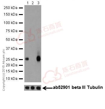
![Western blot - Anti-PHD3 antibody [EPR17869] (ab184714)](http://img.lianshimall.com/statics/attachment/goods/pl20160426/abcamMainImgPrimary/detail/ab18/ab184714WBe.jpg)
Western blot - Anti-PHD3 antibody [EPR17869] (ab184714)
All lanes : Anti-PHD3 antibody [EPR17869] (ab184714) at 1/2000 dilution
Lane 1 : Mouse kidney lysate
Lane 2 : Mouse spleen lysate
Lane 3 : Rat brain lysate
Lane 4 : RAW 264.7 (Mouse macrophage cells transformed with Abelson murine leukemia virus) whole cell lysate
Lane 5 : PC-12 (Rat adrenal gland pheochromocytoma) whole cell lysate
Lane 6 : NIH/3T3 (Mouse embyro fibroblast cells) whole cell lysate
Lysates/proteins at 10 µg per lane.
Secondary
Goat Anti-Rabbit IgG, (H+L),Peroxidase conjugated at 1/1000 dilution
Predicted band size : 27 kDa
Observed band size : 27 kDa
Exposure time : 15 secondsBlocking/Dilution buffer: 5% NFDM/TBST.
![Western blot - Anti-PHD3 antibody [EPR17869] (ab184714)](http://img.lianshimall.com/statics/attachment/goods/pl20160426/abcamMainImgPrimary/detail/ab18/ab184714WBf.jpg)
Western blot - Anti-PHD3 antibody [EPR17869] (ab184714)
All lanes : Anti-PHD3 antibody [EPR17869] (ab184714) at 1/2000 dilution
Lane 1 : Untreated MCF7 (Human breast adenocarcinoma cell line) whole cell lysate
Lane 2 : MCF7 cell lysate treated with 0.5mM CoCl2 (Cobalt (II) chloride) for 6 hours
Lysates/proteins at 10 µg per lane.
Secondary
Goat Anti-Rabbit IgG, (H+L), Peroxidase conjugated at 1/1000 dilution
Predicted band size : 27 kDa
Observed band size : 27 kDa
Exposure time : 1 minuteBlocking/Dilution buffer: 5% NFDM/TBST.
PHD3 expression was induced by CoCl2 treatment (PMID: 18337469).
![Immunocytochemistry/ Immunofluorescence - Anti-PHD3 antibody [EPR17869] (ab184714)](http://img.lianshimall.com/statics/attachment/goods/pl20160426/abcamMainImgPrimary/detail/ab18/ab184714IFa.jpg)
Immunocytochemistry/ Immunofluorescence - Anti-PHD3 antibody [EPR17869] (ab184714)
Immunofluorescent analysis of 4% paraformaldehyde-fixed, 0.1% Triton X-100 permeabilized A549 (Human lung carcinoma) cells labeling PHD3 with ab184714 at 1/250 dilution, followed by Goat anti-rabbit IgG (Alexa Fluor® 488) (ab150077) secondary antibody at 1/1000 dilution (green).
Confocal image showing weakly cytoplasm and nuclear staining on A549 cell line.
The nuclear counterstain is DAPI (blue).
Tubulin is detected with ab7291 (anti-Tubulin mouse mAb) at 1/1000 dilution and ab150120 (AlexaFluor®594 Goat anti-Mouse secondary) at 1/1000 dilution (red).
The negative controls are as follows:-
-ve control 1: ab184714 at 1/250 dilution followed by ab150120 (AlexaFluor®594 Goat anti-Mouse secondary) at 1/1000 dilution.
-ve control 2: ab7291 (anti-Tubulin mouse mAb) at 1/1000 dilution followed by ab150077 (Alexa Fluor®488 Goat Anti-Rabbit IgG H&L) at 1/1000 dilution.
![Immunocytochemistry/ Immunofluorescence - Anti-PHD3 antibody [EPR17869] (ab184714)](http://img.lianshimall.com/statics/attachment/goods/pl20160426/abcamMainImgPrimary/detail/ab18/ab184714IFb.jpg)
Immunocytochemistry/ Immunofluorescence - Anti-PHD3 antibody [EPR17869] (ab184714)
Immunofluorescent analysis of 4% paraformaldehyde-fixed, 0.1% Triton X-100 permeabilized PC-12 (Rat adrenal gland pheochromocytoma) cells labeling PHD3 with ab184714 at 1/250 dilution, followed by Goat anti-rabbit IgG (Alexa Fluor® 488) (ab150077) secondary antibody at 1/1000 dilution (green).
Confocal image showing weakly cytoplasm and nuclear staining on PC-12 cells.
The nuclear counterstain is DAPI (blue).
Tubulin is detected with ab7291 (anti-Tubulin mouse mAb) at 1/1000 dilution and ab150120 (AlexaFluor®594 Goat anti-Mouse secondary) at 1/1000 dilution (red).
The negative controls are as follows:-
-ve control 1: ab184714 at 1/250 dilution followed by ab150120 (AlexaFluor®594 Goat anti-Mouse secondary) at 1/1000 dilution.
-ve control 2: ab7291 (anti-Tubulin mouse mAb) at 1/1000 dilution followed by ab150077 (Alexa Fluor®488 Goat Anti-Rabbit IgG H&L) at 1/1000 dilution.
![Immunoprecipitation - Anti-PHD3 antibody [EPR17869] (ab184714)](http://img.lianshimall.com/statics/attachment/goods/pl20160426/abcamMainImgPrimary/detail/ab18/ab1847144IP.jpg)
Immunoprecipitation - Anti-PHD3 antibody [EPR17869] (ab184714)
PHD3 was immunoprecipitated from 1mg of NIH/3T3 (Mouse embyro fibroblast cells) whole cell lysate with ab184714 at 1/70 dilution.
Western blot was performed from the immunoprecipitate using ab184714 at 1/1000 dilution.
VeriBlot for IP secondary antibody (HRP) (ab131366) was used as secondary antibody at 1/10000 dilution.
Lane 1: NIH/3T3 whole cell lysate 10ug (Input).
Lane 2: ab184714 IP in NIH/3T3 whole cell lysate.
Lane 3: NIH/3T3 whole cell lysate supernatant after capture (unbound).
Lane 4: Rabbit monoclonal IgG (ab172730) instead of ab184714 in NIH/3T3 whole cell lysate.
Blocking and dilution buffer and concentration: 5% NFDM/TBST.
Exposure time: 30 seconds.
ab184714 is not a strong binder for IP - only a partial amount of the target protein in the lysate was immune-precipitated.


 粤公网安备44196802000105号