


详细说明
概述
产品名称Anti-PIGK抗体[EPR17843]
描述
兔单克隆抗体[EPR17843] to PIGK
经测试应用ICC/IF,Flow Cyt,IP,WB
种属反应性
与反应: Mouse, Rat, Human
免疫原
Recombinant fragment within Human PIGK aa 100 to the C-terminus. The exact sequence is proprietary.
Database link: Q92643Run BLAST with
 Run BLAST with
阳性对照
WB: HT1080, HEK-293 and C6 cell lysates; Human pancreas and fetal liver lysates; Mouse brain, mouse heart, mouse kidney, mouse spleen, rat brain and rat kidney lysates. ICC/IF: A549 and Jurkat cells. Flow Cyt: HEK-293 cells. IP: HEK-293 whole cell lysate.
常规说明
This product is a recombinant rabbit monoclonal antibody.
Produced using Abcam’s RabMAb® technology. RabMAb® technology is covered by the following U.S. Patents, No. 5,675,063 and/or 7,429,487.
性能
形式Liquid
存放说明Shipped at 4°C. Store at +4°C short term (1-2 weeks). Upon delivery aliquot. Store at -20°C long term. Avoid freeze / thaw cycle.
存储溶液Preservative: 0.01% Sodium azide
Constituents: 59% PBS, 40% Glycerol, 0.05% BSA纯度Protein A purified
克隆单克隆
克隆编号EPR17843
同种型IgG
研究领域
Signal Transduction
Protein Trafficking
ER Proteins
Anti-PIGK antibody [EPR17843] 图像
![Immunocytochemistry/ Immunofluorescence - Anti-PIGK antibody [EPR17843] (ab201693)](http://img.lianshimall.com/statics/attachment/goods/pl20160426/abcamMainImgPrimary/detail/ab2/ab201693kat.jpg)
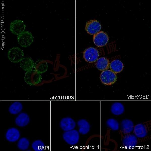
Immunocytochemistry/ Immunofluorescence - Anti-PIGK antibody [EPR17843] (ab201693)
Immunofluorescent analysis of 4% paraformaldehyde-fixed, 0.1% Triton X-100 permeabilized Jurkat (Human T cell leukemia cells from peripheral blood) cells labeling PIGK with ab201693 at 1/500 dilution, followed by Goat anti-rabbit IgG (Alexa Fluor® 488) (ab150077) secondary antibody at 1/500 dilution (green).
Confocal image showing cytoplasmic staining on Jurkat cell line.
The nuclear counter stain is DAPI (blue).
Tubulin is detected with ab7291 (anti-Tubulin mouse mAb) at 1/1000 dilution and ab150120 (AlexaFluor®594 Goat anti-Mouse secondary) at 1/500 dilution (red).
The negative controls are as follows:
-ve control 1: ab201693 at 1/1000 dilution followed by ab150120 (AlexaFluor®594 Goat anti-Mouse secondary) at 1/500 dilution.
-ve control 2: ab7291 (anti-Tubulin mouse mAb) at 1/1000 dilution followed by ab150077 (Alexa Fluor®488 Goat Anti-Rabbit IgG H&L) at 1/500 dilution.![Immunocytochemistry/ Immunofluorescence - Anti-PIGK antibody [EPR17843] (ab201693)](http://img.lianshimall.com/statics/attachment/goods/pl20160426/abcamMainImgPrimary/detail/ab2/ab201693549.jpg)
Immunocytochemistry/ Immunofluorescence - Anti-PIGK antibody [EPR17843] (ab201693)
Immunofluorescent analysis of 100% methanol-fixed, 0.1% Triton X-100 permeabilized A549 (Human lung carcinoma) cells labeling PIGK with ab201693 at 1/500 dilution, followed by Goat anti-rabbit IgG (Alexa Fluor® 488) (ab150077) secondary antibody at 1/500 dilution (green).
Confocal image showing cytoplasmic staining on A549 cell line.
The nuclear counter stain is DAPI (blue).
Tubulin is detected with ab7291 (anti-Tubulin mouse mAb) at 1/1000 dilution and ab150120 (AlexaFluor®594 Goat anti-Mouse secondary) at 1/500 dilution (red).
The negative controls are as follows:
-ve control 1: ab201693 at 1/1000 dilution followed by ab150120 (AlexaFluor®594 Goat anti-Mouse secondary) at 1/500 dilution.
-ve control 2: ab7291 (anti-Tubulin mouse mAb) at 1/1000 dilution followed by ab150077 (Alexa Fluor®488 Goat Anti-Rabbit IgG H&L) at 1/500 dilution.![Western blot - Anti-PIGK antibody [EPR17843] (ab201693)](http://img.lianshimall.com/statics/attachment/goods/pl20160426/abcamMainImgPrimary/detail/ab2/ab201693WBa.jpg)
Western blot - Anti-PIGK antibody [EPR17843] (ab201693)
All lanes : Anti-PIGK antibody [EPR17843] (ab201693) at 1/2000 dilution
Lane 1 : HT1080 (Human fibrosarcoma cells) cell lysate
Lane 2 : HEK-293 (Human epithelial cells from embryonic kidney) cell lysate
Lysates/proteins at 20 µg per lane.
Secondary
Goat Anti-Rabbit IgG, (H+L), Peroxidase conjugated at 1/1000 dilution
Predicted band size : 45 kDa
Observed band size : 45 kDa
Exposure time : 1 minuteBlocking/Dilution buffer: 5% NFDM/TBST.
![Western blot - Anti-PIGK antibody [EPR17843] (ab201693)](http://img.lianshimall.com/statics/attachment/goods/pl20160426/abcamMainImgPrimary/detail/ab2/ab201693WBb.jpg)
Western blot - Anti-PIGK antibody [EPR17843] (ab201693)
All lanes : Anti-PIGK antibody [EPR17843] (ab201693) at 1/2000 dilution
Lane 1 : Human pancreas lysate
Lane 2 : Human fetal liver lysate
Lysates/proteins at 10 µg per lane.
Secondary
Anti-Rabbit IgG (HRP), specific to the non-reduced form of IgG at 1/1000 dilution
Predicted band size : 45 kDa
Observed band size : 45 kDa
Exposure time : 3 minutesBlocking/Dilution buffer: 5% NFDM/TBST.
![Western blot - Anti-PIGK antibody [EPR17843] (ab201693)](http://img.lianshimall.com/statics/attachment/goods/pl20160426/abcamMainImgPrimary/detail/ab2/ab201693WBc.jpg)
Western blot - Anti-PIGK antibody [EPR17843] (ab201693)
All lanes : Anti-PIGK antibody [EPR17843] (ab201693) at 1/2000 dilution
Lane 1 : Mouse brain lysate
Lane 2 : Mouse heart lysate
Lane 3 : Mouse kidney lysate
Lane 4 : Mouse spleen lysate
Lane 5 : Rat brain lysate
Lane 6 : Rat kidney lysate
Lane 7 : C6 (Rat glial tumor cells) cell lysate
Lysates/proteins at 10 µg per lane.
Secondary
Goat Anti-Rabbit IgG, (H+L), Peroxidase conjugated at 1/1000 dilution
Predicted band size : 45 kDa
Observed band size : 45 kDa
Exposure time : 1 minuteBlocking/Dilution buffer: 5% NFDM/TBST.
![Flow Cytometry - Anti-PIGK antibody [EPR17843] (ab201693)](http://img.lianshimall.com/statics/attachment/goods/pl20160426/abcamMainImgPrimary/detail/ab2/ab2016933FC.jpg)
Flow Cytometry - Anti-PIGK antibody [EPR17843] (ab201693)
Flow cytometric analysis of 2% paraformaldehyde-fixed HEK-293 (Human epithelial cells from embryonic kidney) cells labeling PIGK with ab201693 at 1/100 dilution (red) compared with a rabbit monoclonal IgG isotype control (ab172730; black) and an unlabelled control (cells without incubation with primary antibody and secondary antibody; blue). Goat anti rabbit IgG (FITC) at 1/150 dilution was used as the secondary antibody.
![Immunoprecipitation - Anti-PIGK antibody [EPR17843] (ab201693)](http://img.lianshimall.com/statics/attachment/goods/pl20160426/abcamMainImgPrimary/detail/ab2/ab2016933IP.jpg)
Immunoprecipitation - Anti-PIGK antibody [EPR17843] (ab201693)
PIGK was immunoprecipitated from 1mg of HEK293 (Human epithelial cells from embryonic kidney) whole cell lysate with ab201693 at 1/30 dilution.
Western blot was performed from the immunoprecipitate using ab201693 at 1/2000 dilution.
VeriBlot for IP secondary antibody (HRP) (ab131366) was used as secondary antibody at 1/1500 dilution.
Lane 1: HEK293 whole cell lysate10 µg (Input).
Lane 2: ab201693 IP in HEK293 whole cell lysate.
Lane 3: Rabbit monoclonal IgG (ab172730) instead of ab201693 in HEK293 whole cell lysate.
Blocking and dilution buffer and concentration: 5% NFDM/TBST.
Exposure time: 5 seconds.


 粤公网安备44196802000105号