


详细说明
概述
产品名称Anti-PMM1抗体[EPR17844]
描述
兔单克隆抗体[EPR17844] to PMM1
经测试应用IHC-P,WB
种属反应性
与反应: Mouse, Rat, Human
免疫原
Recombinant fragment within Human PMM1 aa 50 to the C-terminus. The exact sequence is proprietary.
Database link: Q92871Run BLAST with
 Run BLAST with
阳性对照
WB: Human fetal liver, fetal brain, fetal heart and fetal kidney lysates; HepG2, 293, C6, NIH/3T3 and PC-12 whole cell lysates; Mouse brain and rat brain lysates. IHC-P: Human liver, mouse cerebral cortex and rat pancreas tissues.
常规说明
This product is a recombinant rabbit monoclonal antibody.
Produced using Abcam’s RabMAb® technology. RabMAb® technology is covered by the following U.S. Patents, No. 5,675,063 and/or 7,429,487.
性能
形式Liquid
存放说明Shipped at 4°C. Store at +4°C short term (1-2 weeks). Upon delivery aliquot. Store at -20°C long term. Avoid freeze / thaw cycle.
存储溶液Preservative: 0.01% Sodium azide
Constituents: 59% PBS, 40% Glycerol, 0.05% BSA纯度Protein A purified
克隆单克隆
克隆编号EPR17844
同种型IgG
研究领域
Metabolism
Pathways and Processes
Metabolic signaling pathways
Energy transfer pathways
Energy Metabolism
Signal Transduction
Metabolism
Energy Metabolism
Anti-PMM1 antibody [EPR17844] 图像
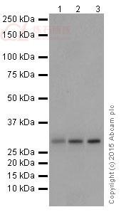
![Western blot - Anti-PMM1 antibody [EPR17844] (ab202058)](http://img.lianshimall.com/statics/attachment/goods/pl20160426/abcamMainImgPrimary/detail/ab2/ab202058b-1.jpg)
Western blot - Anti-PMM1 antibody [EPR17844] (ab202058)
All lanes : Anti-PMM1 antibody [EPR17844] (ab202058) at 1/5000 dilution
Lane 1 : Human fetal liver lysate
Lane 2 : HepG2 (Human liver hepatocellular carcinoma) whole cell lysate
Lane 3 : 293 (Human epithelial cells from embryonic kidney) whole cell lysate
Lysates/proteins at 10 µg per lane.
Secondary
Anti-Rabbit IgG (HRP), specific to the non-reduced form of IgG at 1/1000 dilution
Predicted band size : 30 kDa
Observed band size : 30 kDa
Exposure time : 1 minuteBlocking/Dilution buffer: 5% NFDM/TBST.
![Western blot - Anti-PMM1 antibody [EPR17844] (ab202058)](http://img.lianshimall.com/statics/attachment/goods/pl20160426/abcamMainImgPrimary/detail/ab2/ab202058b-2.jpg)
Western blot - Anti-PMM1 antibody [EPR17844] (ab202058)
All lanes : Anti-PMM1 antibody [EPR17844] (ab202058) at 1/2000 dilution
Lane 1 : Human fetal brain lysate
Lane 2 : Human fetal heart lysate
Lane 3 : Human fetal kidney lysate
Lysates/proteins at 10 µg per lane.
Secondary
Anti-Rabbit IgG (HRP), specific to the non-reduced form of IgG at 1/1000 dilution
Predicted band size : 30 kDa
Observed band size : 30 kDa
Exposure time : 3 minutesBlocking/Dilution buffer: 5% NFDM/TBST.
![Western blot - Anti-PMM1 antibody [EPR17844] (ab202058)](http://img.lianshimall.com/statics/attachment/goods/pl20160426/abcamMainImgPrimary/detail/ab2/ab202058b-3.jpg)
Western blot - Anti-PMM1 antibody [EPR17844] (ab202058)
All lanes : Anti-PMM1 antibody [EPR17844] (ab202058) at 1/2000 dilution
Lane 1 : Mouse brain lysate
Lane 2 : Rat brain lysate
Lysates/proteins at 10 µg per lane.
Secondary
Goat Anti-Rabbit IgG, (H+L), Peroxidase conjugated at 1/1000 dilution
Predicted band size : 30 kDa
Observed band size : 30 kDa
Exposure time : 30 secondsBlocking/Dilution buffer: 5% NFDM/TBST.
![Western blot - Anti-PMM1 antibody [EPR17844] (ab202058)](http://img.lianshimall.com/statics/attachment/goods/pl20160426/abcamMainImgPrimary/detail/ab2/ab202058b-4.jpg)
Western blot - Anti-PMM1 antibody [EPR17844] (ab202058)
All lanes : Anti-PMM1 antibody [EPR17844] (ab202058) at 1/5000 dilution
Lane 1 : C6 (Rat glial tumor cells) whole cell lysate
Lane 2 : PC-12 (Rat adrenal gland pheochromocytoma) whole cell lysate
Lane 3 : NIH/3T3 (Mouse embyro fibroblast cells) whole cell lysate
Lysates/proteins at 10 µg per lane.
Secondary
Goat Anti-Rabbit IgG, (H+L), Peroxidase conjugated at 1/1000 dilution
Predicted band size : 30 kDa
Observed band size : 30 kDa
Exposure time : 30 secondsBlocking/Dilution buffer: 5% NFDM/TBST.
![Immunohistochemistry (Formalin/PFA-fixed paraffin-embedded sections) - Anti-PMM1 antibody [EPR17844] (ab202058)](http://img.lianshimall.com/statics/attachment/goods/pl20160426/abcamMainImgPrimary/detail/ab2/ab202058c-1.jpg)
Immunohistochemistry (Formalin/PFA-fixed paraffin-embedded sections) - Anti-PMM1 antibody [EPR17844] (ab202058)
Immunohistochemical analysis of paraffin-embedded Human liver tissue labeling PMM1 with ab202058 at 1/100 dilution, followed by Goat Anti-Rabbit IgG H&L (HRP) (ab97051) secondary antibody at 1/500 dilution. Cytoplasmic staining on Human liver tissue is observed. Counter stained with Hematoxylin.
Secondary antibody only control: Used PBS instead of primary antibody, secondary antibody is Goat Anti-Rabbit IgG H&L (HRP) (ab97051) at 1/500 dilution.
![Immunohistochemistry (Formalin/PFA-fixed paraffin-embedded sections) - Anti-PMM1 antibody [EPR17844] (ab202058)](http://img.lianshimall.com/statics/attachment/goods/pl20160426/abcamMainImgPrimary/detail/ab2/ab202058c-2.jpg)
Immunohistochemistry (Formalin/PFA-fixed paraffin-embedded sections) - Anti-PMM1 antibody [EPR17844] (ab202058)
Immunohistochemical analysis of paraffin-embedded Mouse cerebral cortex tissue labeling PMM1 with ab202058 at 1/100 dilution, followed by Goat Anti-Rabbit IgG H&L (HRP) (ab97051) secondary antibody at 1/500 dilution. Cytoplasmic staining on mouse cerebral cortex is observed. Counter stained with Hematoxylin.
Secondary antibody only control: Used PBS instead of primary antibody, secondary antibody is Goat Anti-Rabbit IgG H&L (HRP) (ab97051) at 1/500 dilution.
![Immunohistochemistry (Formalin/PFA-fixed paraffin-embedded sections) - Anti-PMM1 antibody [EPR17844] (ab202058)](http://img.lianshimall.com/statics/attachment/goods/pl20160426/abcamMainImgPrimary/detail/ab2/ab202058c-3.jpg)
Immunohistochemistry (Formalin/PFA-fixed paraffin-embedded sections) - Anti-PMM1 antibody [EPR17844] (ab202058)
Immunohistochemical analysis of paraffin-embedded rat pancreas tissue labeling PMM1 with ab202058 at 1/100 dilution, followed by Goat Anti-Rabbit IgG H&L (HRP) (ab97051) secondary antibody at 1/500 dilution. Cytoplasmic staining on rat pancreas is observed. Counter stained with Hematoxylin.
Secondary antibody only control: Used PBS instead of primary antibody, secondary antibody is Goat Anti-Rabbit IgG H&L (HRP) (ab97051) at 1/500 dilution.


 粤公网安备44196802000105号