


详细说明
概述
产品名称Anti-BRSK1抗体[EPR18190]
描述
兔单克隆抗体[EPR18190] to BRSK1
经测试应用ICC/IF,WB
种属反应性
与反应: Mouse, Rat, Human
免疫原
Recombinant fragment within Human BRSK1 aa 350-600. The exact sequence is proprietary.
Database link: Q8TDC3Run BLAST with
 Run BLAST with
阳性对照
WB: SH-SY5Y, Jurkat, PC-12 and NIH/3T3 whole cell lysates; mouse testis lysate; rat brain lysate. ICC/IF: Neuro-2a and NIH/3T3 cells.
??规说明
This product is a recombinant rabbit monoclonal antibody.
Produced using Abcam’s RabMAb® technology. RabMAb® technology is covered by the following U.S. Patents, No. 5,675,063 and/or 7,429,487.
性能
形式Liquid
存放说明Shipped at 4°C. Store at +4°C short term (1-2 weeks). Upon delivery aliquot. Store at -20°C long term. Avoid freeze / thaw cycle.
存储溶液Preservative: 0.01% Sodium azide
Constituents: 59% PBS, 40% Glycerol, 0.05% BSA纯度Protein A purified
克隆单克隆
克隆编号EPR18190
同种型IgG
研究领域
Cell Biology
Cell Cycle
Kinases/Phosphatases
Other
Anti-BRSK1 antibody [EPR18190] 图像
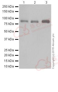
![Western blot - Anti-BRSK1 antibody [EPR18190] (ab206298)](http://img.lianshimall.com/statics/attachment/goods/pl20160426/abcamMainImgPrimary/detail/ab2/ab206298WBa.jpg)
Western blot - Anti-BRSK1 antibody [EPR18190] (ab206298)
All lanes : Anti-BRSK1 antibody [EPR18190] (ab206298) at 1/1000 dilution
Lane 1 : SH-SY5Y (Human neuroblastoma from bone marrow cells) whole cell lysate
Lane 2 : Jurkat (Human T cell leukemia cells from peripheral blood) whole cell lysate
Lane 3 : Mouse testis lysate
Lysates/proteins at 20 µg per lane.
Secondary
Goat Anti-Rabbit IgG H&L (HRP) (ab97051) at 1/1000 dilution
Predicted band size : 85 kDa
Observed band size : 85 kDa
Exposure time : 3 minutesBlocking/Dilution buffer: 5% NFDM/TBST
![Western blot - Anti-BRSK1 antibody [EPR18190] (ab206298)](http://img.lianshimall.com/statics/attachment/goods/pl20160426/abcamMainImgPrimary/detail/ab2/ab206298WBb.jpg)
Western blot - Anti-BRSK1 antibody [EPR18190] (ab206298)
All lanes : Anti-BRSK1 antibody [EPR18190] (ab206298) at 1/1000 dilution
Lane 1 : Rat brain lysate
Lane 2 : Rat heart lysate
Lane 3 : Rat kidney lysate
Lane 4 : Rat spleen lysate
Lysates/proteins at 10 µg per lane.
Secondary
Goat Anti-Rabbit IgG H&L (HRP) (ab97051) at 1/10000 dilution
Predicted band size : 85 kDa
Observed band size : 85 kDa
Exposure time : 3 minutesBlocking/Dilution buffer: 5% NFDM/TBST
![Western blot - Anti-BRSK1 antibody [EPR18190] (ab206298)](http://img.lianshimall.com/statics/attachment/goods/pl20160426/abcamMainImgPrimary/detail/ab2/ab206298WBc.jpg)
Western blot - Anti-BRSK1 antibody [EPR18190] (ab206298)
All lanes : Anti-BRSK1 antibody [EPR18190] (ab206298) at 1/1000 dilution
Lane 1 : PC-12 (Rat adrenal gland pheochromocytoma) whole cell lysate
Lane 2 : NIH/3T3 (Mouse embyro fibroblast cells) whole cell lysate
Lysates/proteins at 10 µg per lane.
Secondary
Goat Anti-Rabbit IgG H&L (HRP) (ab97051) at 1/10000 dilution
Predicted band size : 85 kDa
Observed band size : 85 kDa
Exposure time: Lane 1 - 30 seconds; Lane 2 - 3 minutes.
Blocking/Dilution buffer: 5% NFDM/TBST.
![Immunocytochemistry/ Immunofluorescence - Anti-BRSK1 antibody [EPR18190] (ab206298)](http://img.lianshimall.com/statics/attachment/goods/pl20160426/abcamMainImgPrimary/detail/ab2/ab206298984.jpg)
Immunocytochemistry/ Immunofluorescence - Anti-BRSK1 antibody [EPR18190] (ab206298)
Immunofluorescent analysis of 4% paraformaldehyde-fixed, 0.1% Triton X-100 permeabilized Neuro-2a (Mouse neuroblastoma cells) cells labeling BRSK1 with ab206298 at 1/100 dilution, followed by Goat anti-rabbit IgG (Alexa Fluor® 488) (ab150077) secondary antibody at 1/1000 dilution (green). Confocal image showing both nuclear and cytoplasmic staining on Neuro-2a cell line. The nuclear counter stain is DAPI (blue). Tubulin is detected with ab7291 (anti-Tubulin mouse mAb) at 1/1000 dilution and ab150120 (AlexaFluor®594 Goat anti-Mouse secondary) at 1/1000 dilution (red).
The negative controls are as follows;
-ve control 1: ab206298 at 1/100 dilution followed by ab150120 (AlexaFluor®594 Goat anti-Mouse secondary) at 1/1000 dilution.
-ve control 2: ab7291 (anti-Tubulin mouse mAb) at 1/1000 dilution followed by ab150077 (Alexa Fluor®488 Goat Anti-Rabbit IgG H&L) at 1/1000 dilution![Immunocytochemistry/ Immunofluorescence - Anti-BRSK1 antibody [EPR18190] (ab206298)](http://img.lianshimall.com/statics/attachment/goods/pl20160426/abcamMainImgPrimary/detail/ab2/ab206298985.jpg)
Immunocytochemistry/ Immunofluorescence - Anti-BRSK1 antibody [EPR18190] (ab206298)
Immunofluorescent analysis of 4% paraformaldehyde-fixed, 0.1% Triton X-100 permeabilized NIH/3T3 (Mouse embyro fibroblast cells) cells labeling BRSK1 with ab206298 at 1/100 dilution, followed by Goat anti-rabbit IgG (Alexa Fluor® 488) (ab150077) secondary antibody at 1/1000 dilution (green). Confocal image showing both nuclear and cytoplasmic staining on NIH/3T3 cell line. The nuclear counter stain is DAPI (blue). Tubulin is detected with ab7291 (anti-Tubulin mouse mAb) at 1/1000 dilution and ab150120 (AlexaFluor®594 Goat anti-Mouse secondary) at 1/1000 dilution (red).
The negative controls are as follows;
-ve control 1: ab206298 at 1/100 dilution followed by ab150120 (AlexaFluor®594 Goat anti-Mouse secondary) at 1/1000 dilution.
-ve control 2: ab7291 (anti-Tubulin mouse mAb) at 1/1000 dilution followed by ab150077 (Alexa Fluor®488 Goat Anti-Rabbit IgG H&L) at 1/1000 dilution


 粤公网安备44196802000105号