


详细说明
概述
产品名称Anti-Caspase-9抗体[EPR18107]
描述
兔单克隆抗体[EPR18107] to Caspase-9
经测试应用IHC-P,WB,ICC/IF,IP
种属反应性
与反应: Mouse, Human
免疫原
Recombinant fragment within Human Caspase-9 aa 100-300. The exact sequence is proprietary.
Database link: P55211Run BLAST with
 Run BLAST with
阳性对照
WB: HeLa and C2C12 whole cell lysates; Human fetal brain, fetal heart, fetal kidney and fetal liver lysates. IHC-P: Human cervix carcinoma tissue. ICC/IF: HeLa cells. IP: HeLa treated with staurosporine 1uM for 4 hours whole cell lysate.
常规说明
This product is a recombinant rabbit monoclonal antibody.
Produced using Abcam’s RabMAb® technology. RabMAb® technology is covered by the following U.S. Patents, No. 5,675,063 and/or 7,429,487.
性能
形式Liquid
存放说明Shipped at 4°C. Store at +4°C short term (1-2 weeks). Upon delivery aliquot. Store at -20°C long term. Avoid freeze / thaw cycle.
存储溶液Preservative: 0.01% Sodium azide
Constituents: 59% PBS, 40% Glycerol, 0.05% BSA纯度Protein A purified
克隆单克隆
克隆编号EPR18107
同种型IgG
研究领域
Cancer
Cell Death
Apoptosis
Metabolism
Cancer
Cell Death
Apoptosis
Apoptotic Markers
Cytochrome C
Cancer
Cell Death
Apoptosis
Apoptotic Markers
Caspases
Metabolism
Pathways and Processes
Metabolism processes
Apoptosis
Cancer
Invasion/microenvironment
Apoptosis
Cytochrome C
Cancer
Invasion/microenvironment
Apoptosis
Caspases
Cell Biology
Apoptosis
Intracellular
Caspases etc
Caspases
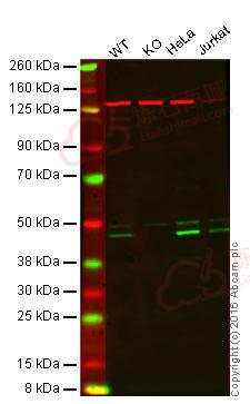
Anti-Caspase-9 antibody [EPR18107] 图像
![Western blot - Anti-Caspase-9 antibody [EPR18107] (ab202068)](http://img.lianshimall.com/statics/attachment/goods/pl20160426/abcamMainImgPrimary/detail/ab2/ab202068OWB.jpg)
Western blot - Anti-Caspase-9 antibody [EPR18107] (ab202068)
Predicted band size : 46 kDa
Lane 1: Wild-type HAP1 cell lysate (20 µg)
Lane 2: Caspase-9 knockout HAP1 cell lysate (20 µg)
Lane 3: HeLa cell lysate (20 µg)
Lane 4: Jurkat cell lysate (20 µg)
Lanes 1 - 4: Merged signal (red and green). Green - ab202068 observed at 46 kDa. Red - loading control, ab8245, observed at 37 kDa.
ab202068 was shown to recognize Caspase-9 when Caspase-9 knockout samples were used, along with additional cross-reactive bands. Wild-type and Caspase-9 knockout samples were subjected to SDS-PAGE. ab202068 and ab8245 (loading control to GAPDH) were diluted at 1/2000 and 1/10 000 respectively and incubated overnight at 4°C. Blots were developed with goat anti-rabbit IgG (H + L) and goat anti-mouse IgG (H + L) secondary antibodies at 1/10 000 dilution for 1 h at room temperature before imaging.![Immunocytochemistry - Anti-Caspase-9 antibody [EPR18107] (ab202068)](http://img.lianshimall.com/statics/attachment/goods/pl20160426/abcamMainImgPrimary/detail/ab2/ab202068068.jpg)
Immunocytochemistry - Anti-Caspase-9 antibody [EPR18107] (ab202068)
Immunofluorescent analysis of 4% paraformaldehyde-fixed, 0.1% Triton X-100 permeabilized HeLa (Human epithelial cells from cervix adenocarcinoma) cells labeling Caspase-9 with ab202068 at 1/500 dilution, followed by Goat anti-rabbit IgG (Alexa Fluor® 488) (ab150077) secondary antibody at 1/500 dilution (green).
Confocal image showing cytoplasmic and nuclear staining on HeLa cell line. The expression increased after treatment with staurosporine (1uM) for 4 hours.
The nuclear counter stain is DAPI (blue). Tubulin is detected with ab7291 (anti-Tubulin mouse mAb) at 1/1000 dilution and ab150120 (AlexaFluor®594 Goat anti-Mouse secondary) at 1/500 dilution (red).
The negative controls are as follows:
-ve control 1: ab202067 at 1/500 dilution followed by ab150120 (AlexaFluor®594 Goat anti-Mouse secondary) at 1/500 dilution.
-ve control 2: ab7291 (anti-Tubulin mouse mAb) at 1/1000 dilution followed by ab150077 (Alexa Fluor®488 Goat Anti-Rabbit IgG H&L) at 1/500 dilution.![Western blot - Anti-Caspase-9 antibody [EPR18107] (ab202068)](http://img.lianshimall.com/statics/attachment/goods/pl20160426/abcamMainImgPrimary/detail/ab2/ab202068WBa.jpg)
Western blot - Anti-Caspase-9 antibody [EPR18107] (ab202068)
All lanes : Anti-Caspase-9 antibody [EPR18107] (ab202068) at 1/20000 dilution
Lane 1 : Untreated HeLa (Human epithelial cells from cervix adenocarcinoma) whole cell lysate
Lane 2 : HeLa (Human epithelial cells from cervix adenocarcinoma) treated with staurosporine 1uM for 4 hours whole cell lysate
Lysates/proteins at 10 µg per lane.
Secondary
Goat Anti-Rabbit IgG, (H+L),Peroxidase conjugated at 1/1000 dilution
Predicted band size : 46 kDa
Observed band size : 35,37,46 kDa (why is the actual band size different from the predicted?)
Exposure time : 30 secondsBlocking/Dilution buffer: 5% NFDM/TBST.
![Immunohistochemistry (Formalin/PFA-fixed paraffin-embedded sections) - Anti-Caspase-9 antibody [EPR18107] (ab202068)](http://img.lianshimall.com/statics/attachment/goods/pl20160426/abcamMainImgPrimary/detail/ab2/ab202068HCa.jpg)
Immunohistochemistry (Formalin/PFA-fixed paraffin-embedded sections) - Anti-Caspase-9 antibody [EPR18107] (ab202068)
Immunohistochemical analysis of paraffin-embedded Human cervix carcinoma tissue labeling Caspase-9 with ab202068 at 1/300 dilution, followed by Goat Anti-Rabbit IgG H&L (HRP) (ab97051) secondary antibody at 1/500 dilution.
Cytoplasmic and nuclear staining on Human cervix carcinoma tissue is observed.
Counter stained with Hematoxylin.
Secondary antibody only control: Used PBS instead of primary antibody, secondary antibody is Goat Anti-Rabbit IgG H&L (HRP) (ab97051) at 1/500 dilution.
![Western blot - Anti-Caspase-9 antibody [EPR18107] (ab202068)](http://img.lianshimall.com/statics/attachment/goods/pl20160426/abcamMainImgPrimary/detail/ab2/ab202068WBb.jpg)
Western blot - Anti-Caspase-9 antibody [EPR18107] (ab202068)
All lanes : Anti-Caspase-9 antibody [EPR18107] (ab202068) at 1/10000 dilution
Lane 1 : Untreated C2C12 (Mouse myoblast cell line) whole cell lysate
Lane 2 : C2C12 (Mouse myoblast cell line) treated with staurosporine 1uM for 4 hours whole cell lysate
Lysates/proteins at 10 µg per lane.
Secondary
Goat Anti-Rabbit IgG, (H+L),Peroxidase conjugated at 1/1000 dilution
Predicted band size : 46 kDa
Observed band size : 37,39,46 kDa (why is the actual band size different from the predicted?)
Exposure time : 3 minutesBlocking/Dilution buffer: 5% NFDM/TBST.
![Western blot - Anti-Caspase-9 antibody [EPR18107] (ab202068)](http://img.lianshimall.com/statics/attachment/goods/pl20160426/abcamMainImgPrimary/detail/ab2/ab202068WBc.jpg)
Western blot - Anti-Caspase-9 antibody [EPR18107] (ab202068)
All lanes : Anti-Caspase-9 antibody [EPR18107] (ab202068) at 1/2000 dilution
Lane 1 : Human fetal brain lysate
Lane 2 : Human fetal heart lysate
Lysates/proteins at 10 µg per lane.
Secondary
Goat Anti-Rabbit IgG, (H+L), Peroxidase conjugated at 1/1000 dilution
Predicted band size : 46 kDa
Observed band size : 46 kDa
Exposure time : 3 minutesBlocking/Dilution buffer: 5% NFDM/TBST.
![Western blot - Anti-Caspase-9 antibody [EPR18107] (ab202068)](http://img.lianshimall.com/statics/attachment/goods/pl20160426/abcamMainImgPrimary/detail/ab2/ab202068WBd.jpg)
Western blot - Anti-Caspase-9 antibody [EPR18107] (ab202068)
All lanes : Anti-Caspase-9 antibody [EPR18107] (ab202068) at 1/2000 dilution
Lane 1 : Human fetal kidney lysate
Lane 2 : Human fetal liver lysate
Lysates/proteins at 10 µg per lane.
Secondary
Goat Anti-Rabbit IgG, (H+L), Peroxidase conjugated at 1/1000 dilution
Predicted band size : 46 kDa
Observed band size : 46 kDa
Exposure time : 30 secondsBlocking/Dilution buffer: 5% NFDM/TBST.
![Immunoprecipitation - Anti-Caspase-9 antibody [EPR18107] (ab202068)](http://img.lianshimall.com/statics/attachment/goods/pl20160426/abcamMainImgPrimary/detail/ab2/ab202068opy.jpg)
Immunoprecipitation - Anti-Caspase-9 antibody [EPR18107] (ab202068)
Caspase-9 was immunoprecipitated from 1mg of HeLa (Human epithelial cells from cervix adenocarcinoma) treated with staurosporine 1uM for 4 hours whole cell lysate with ab202068 at 1/80 dilution.
Western blot was performed from the immunoprecipitate using ab202068 at 1/1000 dilution.
Anti-Rabbit IgG (HRP), specific to the non-reduced form of IgG was used as secondary antibody at 1/1500 dilution.
Lane 1: HeLa treated with staurosporine 1uM for 4 hours whole cell lysate10 µg (Input).
Lane 2: ab202068 IP in HeLa treated with staurosporine 1uM for 4 hours whole cell lysate.
Lane 3: Rabbit monoclonal IgG (ab172730) instead of ab202068 in HeLa treated with staurosporine 1uM for 4 hours whole cell lysate.
Blocking and dilution buffer and concentration: 5% NFDM/TBST.Exposure time: 3 seconds.


 粤公网安备44196802000105号