


详细说明
概述
产品名称Anti-nNOS (neuronal)抗体[EP1855Y]
描述
兔单克隆抗体[EP1855Y] to nNOS (neuronal)
经测试应用WB,IP,Flow Cyt,IHC-Fr,ICC/IF
种属反应性
与反应: Mouse, Rat, Human, Marmoset (common)
免疫原
Synthetic peptide (the amino acid sequence is considered to be commercially sensitive) corresponding to Human nNOS (neuronal). A synthetic peptide corresponding to residues around serine 1417 of human nNOS (neuronal) protein.
阳性对照
WB: Mouse brain tissue lysate. ICC/IF: A673 cells. Flow Cyt: PC-12 cells. IP: Rat brain tissue lysate.
常规说明
This product is a recombinant rabbit monoclonal antibody.
We are constantly working hard to ensure we provide our customers with best in class antibodies. As a result of this work we are pleased to now offer this antibody in purified format. We are in the process of updating our datasheets. The purified format is designated ‘PUR’ on our product labels. If you have any questions regarding this update, please contact our Scientific Support team.
Produced using Abcam’s RabMAb® technology. RabMAb® technology is covered by the following U.S. Patents, No. 5,675,063 and/or 7,429,487.
Alternative versions available:
Anti-nNOS (neuronal) antibody (Alexa Fluor® 594) [EP1855Y] (ab198516)Anti-nNOS (neuronal) antibody (Alexa Fluor® 647) [EP1855Y] (ab198327)
性能
形式Liquid
存放说明Shipped at 4°C. Store at +4°C short term (1-2 weeks). Upon delivery aliquot. Store at -20°C. Avoid freeze / thaw cycle.
存储溶液pH: 7.20
Preservative: 0.01% Sodium azide
Constituents: 59% PBS, 40% Glycerol, 0.05% BSA纯度Protein A purified
克隆单克隆
克隆编号EP1855Y
同种型IgG
研究领域
Metabolism
Types of disease
Cancer
Metabolism
Pathways and Processes
Metabolism processes
Hypoxia
Cancer
Cancer Metabolism
Response to hypoxia
Neuroscience
Neurotransmission
Nitric Oxide
NOS
Anti-nNOS (neuronal) antibody [EP1855Y] 图像
![Western blot - Anti-nNOS (neuronal) antibody [EP1855Y] (ab76067)](http://img.lianshimall.com/statics/attachment/goods/pl20160426/abcamMainImgPrimary/detail/ab7/ab76067pwb.jpg)
Western blot - Anti-nNOS (neuronal) antibody [EP1855Y] (ab76067)
Anti-nNOS (neuronal) antibody [EP1855Y] (ab76067) at 1/100 dilution (unpurified) + Mouse brain tissue lysate at 10 µg
Secondary
Peroxidase-conjugated goat anti-rabbit IgG (H+L) at 1/1000 dilution
Predicted band size : 161 kDa
Blocking buffer and concentration: 5% NFDM/TBST.
Diluting buffer and concentration: 5% NFDM /TBST.
Western blot - Anti-nNOS (neuronal) antibody [EP1855Y] (ab76067)
Anti-nNOS (neuronal) antibody [EP1855Y] (ab76067) at 1/3000 dilution (purified) + Mouse brain tissue lysate at 10 µg
Secondary
Peroxidase-conjugated goat anti-rabbit IgG (H+L) at 1/1000 dilution
Predicted band size : 161 kDa
Blocking buffer and concentration: 5% NFDM/TBST.
Diluting buffer and concentration: 5% NFDM /TBST.
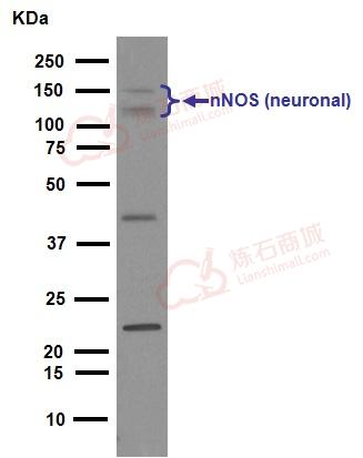
![Western blot - Anti-nNOS (neuronal) antibody [EP1855Y] (ab76067)](http://img.lianshimall.com/statics/attachment/goods/pl20160426/abcamMainImgPrimary/detail/ab7/ab760677_1.JPG)
Western blot - Anti-nNOS (neuronal) antibody [EP1855Y] (ab76067)
Anti-nNOS (neuronal) antibody [EP1855Y] (ab76067) at 1/1000 dilution (unpurified) + Mouse brain tissue lysate at 10 µg
Secondary
HRP-conjugated goat anti-rabbit IgG at 1/2000 dilution
Predicted band size : 161 kDa
Observed band size : 161 kDa
![Immunocytochemistry/ Immunofluorescence - Anti-nNOS (neuronal) antibody [EP1855Y] (ab76067)](http://img.lianshimall.com/statics/attachment/goods/pl20160426/abcamMainImgPrimary/detail/ab7/ab76067cif.jpg)
Immunocytochemistry/ Immunofluorescence - Anti-nNOS (neuronal) antibody [EP1855Y] (ab76067)
Immunocytochemistry/Immunofluorescence analysis of A673 cells labelling nNOS (neuronal) (green) with purified ab76067 at 1/200. Cells were fixed with 4% paraformaldehyde and permeabilized with 0.1% Triton X-100. ab150077, an Alexa Fluor® 488-conjugated goat anti-rabbit IgG (1/500) was used as the secondary antibody. DAPI (blue) was used as the nuclear counterstain.
Control: primary antibody (1/200) and secondary antibody Alexa Fluor® 594-conjugated goat anti-mouse IgG (1/500).
![Immunocytochemistry/ Immunofluorescence - Anti-nNOS (neuronal) antibody [EP1855Y] (ab76067)](http://img.lianshimall.com/statics/attachment/goods/pl20160426/abcamMainImgPrimary/detail/ab7/ab760677-4.jpg)
Immunocytochemistry/ Immunofluorescence - Anti-nNOS (neuronal) antibody [EP1855Y] (ab76067) Image courtesy of Dr Patricia Martin-DeLeon by Abreview.
Unpurified ab76067 staining nNOS (neuronal) in murine sperm cells by Immunocytochemistry/ Immunofluorescence. Cells were fixed with paraformaldehyde and blocked using 2% BSA. Samples were then incubated with undiluted ab76067. The secondary used was a FITC conjugated goat anti-rabbit IgG at a 1/400 dilution.Panel A shows the specific staining of nNOS in sperm while Panel B is the control sample treated with Rabbit IgG.
See Abreview
![Flow Cytometry - nNOS (neuronal) antibody [EP1855Y] (ab76067)](http://img.lianshimall.com/statics/attachment/goods/pl20160426/abcamMainImgPrimary/detail/ab7/ab760677-1.jpg)
Flow Cytometry - nNOS (neuronal) antibody [EP1855Y] (ab76067)
Overlay histogram showing PC-12 cells stained with unpurified ab76067 (red line). The cells were fixed with methanol (5 min) and incubated in 1x PBS / 10% normal goat serum / 0.3M glycine to block non-specific protein-protein interactions. The cells were then incubated with the antibody (unpurified ab76067, 1/50 dilution) for 30 min at 22ºC. The secondary antibody used was DyLight® 488 goat anti-rabbit IgG (H+L) (ab96899) at 1/500 dilution for 30 min at 22ºC. Isotype control antibody (black line) was rabbit monoclonal IgG (1µg/1x106 cells) used under the same conditions. Acquisition of >5,000 events was performed. This antibody gave a slightly decreased signal in PC-12 cells fixed with 4% paraformaldehyde (10 min) used under the same conditions.
Please note that Abcam do not have any data for use of this antibody in non-fixed cells. We welcome any customer feedback.![Flow Cytometry - Anti-nNOS (neuronal) antibody [EP1855Y] (ab76067)](http://img.lianshimall.com/statics/attachment/goods/pl20160426/abcamMainImgPrimary/detail/ab7/ab76067pfc.jpg)
Flow Cytometry - Anti-nNOS (neuronal) antibody [EP1855Y] (ab76067)
Flow cytometry analysis of PC-12 cells labelling nNos (neuronal) with unpurified ab76067 at 1/15 (red). Cells were fixed with 100% methanol. A FITC-conjugated goat anti-rabbit IgG (1/150) was used as the secondary antibody. Green - Isotype control, rabbit monoclonal IgG.
Flow Cytometry - Anti-nNOS (neuronal) antibody [EP1855Y] (ab76067)
Flow cytometry analysis of PC-12 cells labelling nNos (neuronal) with purified ab76067 at 1/600 (red). Cells were fixed with 100% methanol. A FITC-conjugated goat anti-rabbit IgG (1/150) was used as the secondary antibody. Green - Isotype control, rabbit monoclonal IgG.
![Immunoprecipitation - Anti-nNOS (neuronal) antibody [EP1855Y] (ab76067)](http://img.lianshimall.com/statics/attachment/goods/pl20160426/abcamMainImgPrimary/detail/ab7/ab76067pip.jpg)
Immunoprecipitation - Anti-nNOS (neuronal) antibody [EP1855Y] (ab76067)
ab76067 (unpurified) at 1/4 immunoprecipitating nNOS (neuronal) in rat brain tissue lysate. For western blotting, a peroxidase-conjugated goat anti-rabbit IgG (H+L) was used as the secondary antibody (1/1000).
Blocking buffer and concentration: 5% NFDM/TBST.
Diluting buffer and concentration: 5% NFDM /TBST.
Immunoprecipitation - Anti-nNOS (neuronal) antibody [EP1855Y] (ab76067)
ab76067 (purified) at 1/150 immunoprecipitating nNOS (neuronal) in rat brain tissue lysate. For western blotting, a peroxidase-conjugated goat anti-rabbit IgG (H+L) was used as the secondary antibody (1/1000).
Blocking buffer and concentration: 5% NFDM/TBST.
Diluting buffer and concentration: 5% NFDM /TBST.
![Immunohistochemistry (Frozen sections) - Anti-nNOS (neuronal) [EP1855Y] antibody (ab76067)](http://img.lianshimall.com/statics/attachment/goods/pl20160426/abcamMainImgPrimary/detail/ab7/ab76067opy.JPG)
Immunohistochemistry (Frozen sections) - Anti-nNOS (neuronal) [EP1855Y] antibody (ab76067) Image courtesy Anonymous Abreview
IHC-Fr image of nNOS staining on marmoset caudate sections using unpurified ab76067 (1/1000). The tissue was fixed in paraformaldehyde and the sections were then permeabilized using Triton-X. The sections were them blocked using 2% BSA for 2 hour at 20°C. Unpurified ab76067 was diluted 1/1000 and incubated with the sections for 18 hours at 20°C. The secondary antibody used was HRP conjugated goat polyclonal to rabbit IgG (1/1000).
See Abreview


 粤公网安备44196802000105号