


详细说明
概述
产品名称Anti-LSD2 / AOF1抗体[EPR18508]
描述
兔单克隆抗体[EPR18508] to LSD2 / AOF1
经测试应用ICC/IF,IP,WB
种属反应性
与反应: Mouse, Rat, Human
免疫原
Synthetic peptide (the amino acid sequence is considered to be commercially sensitive) within Human LSD2/ AOF1 aa 50-150. The exact sequence is proprietary.
Database link: Q8NB78阳性对照
WB: NIH/3T3, C6, RAW 264.7 and PC-12 cell lysates; Human fetal brain and fetal heart lysates; Mouse thymus lysate. ICC/IF: HeLa and A431 cells. IP: K562 whole cell lysate.
常规说明
This product is a recombinant rabbit monoclonal antibody.
Produced using Abcam’s RabMAb® technology. RabMAb® technology is covered by the following U.S. Patents, No. 5,675,063 and/or 7,429,487.
性能
形式Liquid
存放说明Shipped at 4°C. Store at +4°C short term (1-2 weeks). Upon delivery aliquot. Store at -20°C long term. Avoid freeze / thaw cycle.
存储溶液Preservative: 0.01% Sodium azide
Constituents: 59% PBS, 40% Glycerol, 0.05% BSA纯度Protein A purified
克隆单克隆
克隆编号EPR18508
同种型IgG
研究领域
Epigenetics and Nuclear Signaling
Chromatin Modifying Enzymes
Methylation
Lysine demethylation
Epigenetics and Nuclear Signaling
Chromatin Modifying Enzymes
Methylation
Anti-LSD2 / AOF1 antibody [EPR18508] 图像
![Western blot - Anti-LSD2 / AOF1 antibody [EPR18508] (ab193080)](http://img.lianshimall.com/statics/attachment/goods/pl20160426/abcamMainImgPrimary/detail/ab19/ab193080OWB.jpg)
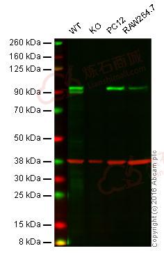
Western blot - Anti-LSD2 / AOF1 antibody [EPR18508] (ab193080)
Predicted band size : 92 kDa
Lane 1: Wild-type HAP1 cell lysate (20 µg)
Lane 2: LSD2 / AOF1 knockout HAP1 cell lysate (20 µg)
Lane 3: PC12 cell lysate (20 µg)
Lane 4: Raw264.7 cell lysate (20 µg)
Lanes 1 - 4: Merged signal (red and green). Green - ab193080 observed at 95 kDa. Red - loading control, ab8245, observed at 37 kDa.
ab193080 was shown to specifically react with LSD2 / AOF1 when LSD2 / AOF1 knockout samples were used. Wild-type and LSD2 / AOF1 knockout samples were subjected to SDS-PAGE. ab193080 and ab8245 (loading control to GAPDH) were diluted at 1/2000 and 1/10 000 respectively and incubated overnight at 4°C. Blots were developed with goat anti-rabbit IgG (H + L) and goat anti-mouse IgG (H + L) secondary antibodies at 1/10 000 dilution for 1 h at room temperature before imaging.![Western blot - Anti-LSD2 / AOF1 antibody [EPR18508] (ab193080)](http://img.lianshimall.com/statics/attachment/goods/pl20160426/abcamMainImgPrimary/detail/ab19/ab193080WBc.jpg)
Western blot - Anti-LSD2 / AOF1 antibody [EPR18508] (ab193080)
All lanes : Anti-LSD2 / AOF1 antibody [EPR18508] (ab193080) at 1/2000 dilution
Lane 1 : Human fetal brain lysate
Lane 2 : Human fetal heart lysate
Lysates/proteins at 10 µg per lane.
Secondary
Anti-Rabbit IgG (HRP), specific to the non-reduced form of IgG at 1/10000 dilution
Predicted band size : 92 kDa
Observed band size : 92 kDa
Exposure time : 3 minutesBlocking/Dilution buffer: 5% NFDM/TBST.
![Western blot - Anti-LSD2 / AOF1 antibody [EPR18508] (ab193080)](http://img.lianshimall.com/statics/attachment/goods/pl20160426/abcamMainImgPrimary/detail/ab19/ab193080WBd.jpg)
Western blot - Anti-LSD2 / AOF1 antibody [EPR18508] (ab193080)
All lanes : Anti-LSD2 / AOF1 antibody [EPR18508] (ab193080) at 1/2000 dilution
Lane 1 : NIH/3T3 (Mouse embyro fibroblast cells) cell lysate
Lane 2 : Mouse thymus lysate
Lysates/proteins at 20 µg per lane.
Secondary
Goat Anti-Rabbit IgG H&L (HRP) (ab97051) at 1/50000 dilution
Predicted band size : 92 kDa
Observed band size : 92 kDa
Exposure time : 3 minutesBlocking/Dilution buffer: 5% NFDM/TBST.
![Western blot - Anti-LSD2 / AOF1 antibody [EPR18508] (ab193080)](http://img.lianshimall.com/statics/attachment/goods/pl20160426/abcamMainImgPrimary/detail/ab19/ab193080WBe.jpg)
Western blot - Anti-LSD2 / AOF1 antibody [EPR18508] (ab193080)
All lanes : Anti-LSD2 / AOF1 antibody [EPR18508] (ab193080) at 1/2000 dilution
Lane 1 : C6 (Rat glial tumor cells) cell lysate
Lane 2 : RAW 264.7 (Mouse macrophage cells transformed with Abelson murine leukemia virus) cell lysate
Lane 3 : PC-12 (Rat adrenal gland pheochromocytoma) cell lysate
Lysates/proteins at 10 µg per lane.
Secondary
Goat Anti-Rabbit IgG H&L (HRP) (ab97051) at 1/1000 dilution
Predicted band size : 92 kDa
Observed band size : 92 kDa
Exposure time : 3 minutesBlocking/Dilution buffer: 5% NFDM/TBST.
![Immunocytochemistry/ Immunofluorescence - Anti-LSD2 / AOF1 antibody [EPR18508] (ab193080)](http://img.lianshimall.com/statics/attachment/goods/pl20160426/abcamMainImgPrimary/detail/ab19/ab193080ged.jpg)
Immunocytochemistry/ Immunofluorescence - Anti-LSD2 / AOF1 antibody [EPR18508] (ab193080)
Immunofluorescent analysis of 4% paraformaldehyde-fixed, 0.1% Triton X-100 permeabilized HeLa (Human epithelial cells from cervix adenocarcinoma) cells labeling LSD2 / AOF1 with ab193080 at 1/500 dilution, followed by Goat anti-rabbit IgG (Alexa Fluor® 488) (ab150077) secondary antibody at 1/1000 dilution (green).
Confocal image showing nuclear staining on HeLa cell line.
The nuclear counterstain is DAPI (blue).
Tubulin is detected with ab7291 (anti-Tubulin mouse mAb) at 1/1000 dilution and ab150120 (AlexaFluor®594 Goat anti-Mouse secondary) at 1/1000 dilution (red).
The negative controls are as follows:
-ve control 1: ab193080 at 1/500 dilution followed by ab150120 (AlexaFluor®594 Goat anti-Mouse secondary) at 1/1000 dilution.
-ve control 2: ab7291 (anti-Tubulin mouse mAb) at 1/1000 dilution followed by ab150077 (Alexa Fluor®488 Goat Anti-Rabbit IgG H&L) at 1/1000 dilution.Immunocytochemistry/ Immunofluorescence - Anti-LSD2 / AOF1 antibody [EPR18508] (ab193080)
Immunofluorescent analysis of 4% paraformaldehyde-fixed, 0.1% Triton X-100 permeabilized A431 (Human epidermoid carcinoma) cells labeling LSD2 / AOF1 with ab193080 at 1/500 dilution, followed by Goat anti-rabbit IgG (Alexa Fluor® 488) (ab150077) secondary antibody at 1/1000 dilution (green).
Confocal image showing nuclear staining on A431 cell line.
The nuclear counterstain is DAPI (blue).
Tubulin is detected with ab7291 (anti-Tubulin mouse mAb) at 1/1000 dilution and ab150120 (AlexaFluor®594 Goat anti-Mouse secondary) at 1/1000 dilution (red).
The negative controls are as follows:-
-ve control 1: ab193080 at 1/500 dilution followed by ab150120 (AlexaFluor®594 Goat anti-Mouse secondary) at 1/1000 dilution.
-ve control 2: ab7291 (anti-Tubulin mouse mAb) at 1/1000 dilution followed by ab150077 (Alexa Fluor®488 Goat Anti-Rabbit IgG H&L) at 1/1000 dilution.![Immunoprecipitation - Anti-LSD2 / AOF1 antibody [EPR18508] (ab193080)](http://img.lianshimall.com/statics/attachment/goods/pl20160426/abcamMainImgPrimary/detail/ab19/ab193080opy.jpg)
Immunoprecipitation - Anti-LSD2 / AOF1 antibody [EPR18508] (ab193080)
LSD2 / AOF1 was immunoprecipitated from 1mg of K562 (Human chronic myelogenous leukemia cells from bone marrow) whole cell lysate with ab193080 at 1/80 dilution.
Lane 1: K562 whole cell lysate 10ug (Input).
Lane 2: ab193080 IP in K562 whole cell lysate.
Lane 3: Rabbit monoclonal IgG (ab172730) instead of ab193080 in K562 whole cell lysate.
Western blot was performed from the immunoprecipitate using ab193080 at 1/1000 dilution. Anti-Rabbit IgG (HRP), specific to the non-reduced form of IgG, was used as secondary antibody at 1/1500.
Blocking and dilution buffer and concentration: 5% NFDM/TBST.
Exposure time: 8 seconds.


 粤公网安备44196802000105号