


详细说明
概述
产品名称Anti-PKC beta 1抗体[EPR18512]
描述
兔单克隆抗体[EPR18512] to PKC beta 1
经测试应用WB,IHC-P,ICC/IF,Flow Cyt
种属反应性
与反应: Mouse, Rat, Human
免疫原
Synthetic peptide (the amino acid sequence is considered to be commercially sensitive) within Human PKC beta 1 aa 600 to the C-terminus. The exact sequence is proprietary.
Database link: P05771阳性对照
WB: Active human PKC beta 1 full length protein; Human fetal brain whole cell lysate; L-929, C2C12, U-87 MG, HeLa, C6 and NIH/3T3 cell lysates; Mouse brain and rat brain lysates. IHC-P: Human colon, Human colon cancer, mouse kidney and rat spleen tissues. ICC/IF: HeLa and K562 cells. Flow Cyt: K562 cells.
常规说明
This product is a recombinant rabbit monoclonal antibody.
Produced using Abcam’s RabMAb® technology. RabMAb® technology is covered by the following U.S. Patents, No. 5,675,063 and/or 7,429,487.
性能
形式Liquid
存放说明Shipped at 4°C. Store at +4°C short term (1-2 weeks). Upon delivery aliquot. Store at -20°C long term. Avoid freeze / thaw cycle.
存储溶液Preservative: 0.01% Sodium azide
Constituents: 59% PBS, 40% Glycerol, 0.05% BSA纯度Protein A purified
克隆单克隆
克隆编号EPR18512
同种型IgG
研究领域
Signal Transduction
Protein Phosphorylation
Ser / Thr Kinases
PKC
Anti-PKC beta 1 antibody [EPR18512] 图像
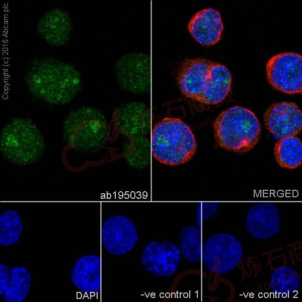
![Immunocytochemistry/ Immunofluorescence - Anti-PKC beta 1 antibody [EPR18512] (ab195039)](http://img.lianshimall.com/statics/attachment/goods/pl20160426/abcamMainImgPrimary/detail/ab19/ab1950399-2.jpg)
Immunocytochemistry/ Immunofluorescence - Anti-PKC beta 1 antibody [EPR18512] (ab195039)
Immunofluorescent analysis of 4% paraformaldehyde-fixed, 0.1% Triton X-100 permeabilized K562 (Human chronic myelogenous leukemia cells from bone marrow) cells labeling PKC beta 1 with ab195039 at 1/100 dilution, followed by Goat anti-rabbit IgG (Alexa Fluor® 488) (ab150077) secondary antibody at 1/1000 dilution (green). Confocal image showing nuclear and cytoplasm staining on K562 cell line. The nuclear counter stain is DAPI (blue). Tubulin is detected with ab7291 (anti-Tubulin mouse mAb) at 1/1000 dilution and ab150120 (AlexaFluor®594 Goat anti-Mouse secondary) at 1/1000 dilution (red).
The negative controls are as follows:
-ve control 1: ab195039 at 1/100 dilution followed by ab150120 (AlexaFluor®594 Goat anti-Mouse secondary) at 1/1000 dilution.
-ve control 2: ab7291 (anti-Tubulin mouse mAb) at 1/1000 dilution followed by ab150077 (Alexa Fluor®488 Goat Anti-Rabbit IgG H&L) at 1/1000 dilution.![Immunocytochemistry/ Immunofluorescence - Anti-PKC beta 1 antibody [EPR18512] (ab195039)](http://img.lianshimall.com/statics/attachment/goods/pl20160426/abcamMainImgPrimary/detail/ab19/ab195039039.jpg)
Immunocytochemistry/ Immunofluorescence - Anti-PKC beta 1 antibody [EPR18512] (ab195039)
Immunofluorescent analysis of 4% paraformaldehyde-fixed, 0.1% Triton X-100 permeabilized HeLa (Human epithelial cells from cervix adenocarcinoma) cells labeling PKC beta 1 with ab195039 at 1/100 dilution, followed by Goat anti-rabbit IgG (Alexa Fluor® 488) (ab150077) secondary antibody at 1/1000 dilution (green). Confocal image showing nuclear and cytoplasm staining on HeLa cell line. The nuclear counter stain is DAPI (blue). Tubulin is detected with ab7291 (anti-Tubulin mouse mAb) at 1/1000 dilution and ab150120 (AlexaFluor®594 Goat anti-Mouse secondary) at 1/1000 dilution (red).
The negative controls are as follows:
-ve control 1: ab195039 at 1/100 dilution followed by ab150120 (AlexaFluor®594 Goat anti-Mouse secondary) at 1/1000 dilution.
-ve control 2: ab7291 (anti-Tubulin mouse mAb) at 1/1000 dilution followed by ab150077 (Alexa Fluor®488 Goat Anti-Rabbit IgG H&L) at 1/1000 dilution.![Western blot - Anti-PKC beta 1 antibody [EPR18512] (ab195039)](http://img.lianshimall.com/statics/attachment/goods/pl20160426/abcamMainImgPrimary/detail/ab19/ab195039b-1.jpg)
Western blot - Anti-PKC beta 1 antibody [EPR18512] (ab195039)
All lanes : Anti-PKC beta 1 antibody [EPR18512] (ab195039) at 1/2000 dilution
Lane 1 : Active human PKC alpha full length protein
Lane 2 : Active human PKC beta 1 full length protein
Lane 3 : Active human PKC beta 2 full length protein
Lane 4 : Active human PKC gamma full length protein
Lane 5 : Active human PKC delta full length protein
Lane 6 : Active human PKC eta full length protein
Lane 7 : Active human PKC epsilon full length protein
Lane 8 : Active human PKC theta full length protein
Lane 9 : Active human PKC mu full length protein
Lysates/proteins at 0.02 µg per lane.
Secondary
Goat Anti-Rabbit IgG H&L (HRP) (ab97051) at 1/50000 dilution
Predicted band size : 77 kDa
Observed band size : 103 kDa (why is the actual band size different from the predicted?)
Exposure time : 1 secondBlocking/Dilution buffer: 5% NFDM/TBST.
Active human PKC alpha full length protein (ab55672) contains aa1-672 with GST-tag; Active human PKC beta 1 full length protein (ab60840) contains aa1-671 with GST-tag; Active human PKC beta 2 full length protein (ab60841) contains aa1-673 with GST-tag; Active human PKC gamma full length protein (ab60842) contains aa1-677 with GST-tag; Active human PKC delta full length protein (ab60844) contains aa1-676 with GST-tag; Active human PKC eta full length protein (ab60849) contains aa1-683 with GST-tag; Active human PKC epsilon full length protein (ab60847) contains aa1-737 with GST-tag; Active human PKC theta full length protein (ab56641) contains aa1-706 with GST-tag; Active human PKC mu full length protein (ab60873) contains aa1-912 with GST-tag.
![Western blot - Anti-PKC beta 1 antibody [EPR18512] (ab195039)](http://img.lianshimall.com/statics/attachment/goods/pl20160426/abcamMainImgPrimary/detail/ab19/ab195039b-2.jpg)
Western blot - Anti-PKC beta 1 antibody [EPR18512] (ab195039)
All lanes : Anti-PKC beta 1 antibody [EPR18512] (ab195039) at 1/10000 dilution
Lane 1 : Human fetal brain whole cell lysate
Lane 2 : L-929 (Mouse connective tissue fibroblast cells) whole cell lysate
Lane 3 : C2C12 (Mouse myoblast cell line) whole cell lysate
Lysates/proteins at 10 µg per lane.
Secondary
Anti-Rabbit IgG (HRP), specific to the non-reduced form of IgG at 1/50000 dilution
Predicted band size : 77 kDa
Observed band size : 40,77 kDa (why is the actual band size different from the predicted?)
Exposure time : 3 minutesBlocking/Dilution buffer: 5% NFDM/TBST.
The peptide immunogen corresponds to the C terminal region of human PKC beta 1. The antibody recognizes the full length (~77KD) and the catalytic domain (~40KD) of PKC beta 1 (Geraldes P and King GL, 2010. Circ Res. 106, 1319-1331.
![Western blot - Anti-PKC beta 1 antibody [EPR18512] (ab195039)](http://img.lianshimall.com/statics/attachment/goods/pl20160426/abcamMainImgPrimary/detail/ab19/ab195039b-3.jpg)
Western blot - Anti-PKC beta 1 antibody [EPR18512] (ab195039)
All lanes : Anti-PKC beta 1 antibody [EPR18512] (ab195039) at 1/10000 dilution
Lane 1 : U-87 MG (Human glioblastoma-astrocytoma epithelial cell line) whole cell lysate
Lane 2 : HeLa (Human epithelial cells from cervix adenocarcinoma) whole cell lysate
Lysates/proteins at 10 µg per lane.
Secondary
Goat Anti-Rabbit IgG H&L (HRP) (ab97051) at 1/50000 dilution
Predicted band size : 77 kDa
Observed band size : 40,77 kDa (why is the actual band size different from the predicted?)
Exposure time : 30 secondsBlocking/Dilution buffer: 5% NFDM/TBST.
The peptide immunogen corresponds to the C terminal region of human PKC beta 1. The antibody recognizes the full length (~77KD) and the catalytic domain (~40KD) of PKC beta 1 (Geraldes P and King GL, 2010. Circ Res. 106, 1319-1331.
![Western blot - Anti-PKC beta 1 antibody [EPR18512] (ab195039)](http://img.lianshimall.com/statics/attachment/goods/pl20160426/abcamMainImgPrimary/detail/ab19/ab195039b-4.jpg)
Western blot - Anti-PKC beta 1 antibody [EPR18512] (ab195039)
All lanes : Anti-PKC beta 1 antibody [EPR18512] (ab195039) at 1/2000 dilution
Lane 1 : Mouse brain lysate
Lane 2 : Rat brain lysate
Lysates/proteins at 10 µg per lane.
Secondary
Goat Anti-Rabbit IgG H&L (HRP) (ab97051) at 1/50000 dilution
Predicted band size : 77 kDa
Observed band size : 40,77 kDa (why is the actual band size different from the predicted?)
Exposure time : 15 secondsBlocking/Dilution buffer: 5% NFDM/TBST.
The peptide immunogen corresponds to the C terminal region of human PKC beta 1. The antibody recognizes the full length (~77KD) and the catalytic domain (~40KD) of PKC beta 1 (Geraldes P and King GL, 2010. Circ Res. 106, 1319-1331.
![Western blot - Anti-PKC beta 1 antibody [EPR18512] (ab195039)](http://img.lianshimall.com/statics/attachment/goods/pl20160426/abcamMainImgPrimary/detail/ab19/ab195039b-5.jpg)
Western blot - Anti-PKC beta 1 antibody [EPR18512] (ab195039)
All lanes : Anti-PKC beta 1 antibody [EPR18512] (ab195039) at 1/2000 dilution
Lane 1 : C6 (Rat glial tumor cells) whole cell lysate
Lane 2 : NIH/3T3 (Mouse embyro fibroblast cells) whole cell lysate
Lysates/proteins at 10 µg per lane.
Secondary
Goat Anti-Rabbit IgG H&L (HRP) (ab97051) at 1/50000 dilution
Predicted band size : 77 kDa
Observed band size : 40,77 kDa (why is the actual band size different from the predicted?)
Exposure time : 3 minutesBlocking/Dilution buffer: 5% NFDM/TBST.
The peptide immunogen corresponds to the C terminal region of human PKC beta 1. The antibody recognizes the full length (~77KD) and the catalytic domain (~40KD) of PKC beta 1 (Geraldes P and King GL, 2010. Circ Res. 106, 1319-1331.
![Immunohistochemistry (Formalin/PFA-fixed paraffin-embedded sections) - Anti-PKC beta 1 antibody [EPR18512] (ab195039)](http://img.lianshimall.com/statics/attachment/goods/pl20160426/abcamMainImgPrimary/detail/ab19/ab195039HCa.jpg)
Immunohistochemistry (Formalin/PFA-fixed paraffin-embedded sections) - Anti-PKC beta 1 antibody [EPR18512] (ab195039)
Immunohistochemical analysis of paraffin-embedded Human colon tissue labeling PKC beta 1 with ab195039 at 1/100 dilution, followed by Goat Anti-Rabbit IgG H&L (HRP) (ab97051) at 1/500 dilution. Nucleus and weak cytoplasm staining on epithelial cells of Human colon is observed. Counter stained with Hematoxylin.
Secondary antibody only control: Used PBS instead of primary antibody, secondary antibody is Goat Anti-Rabbit IgG H&L (HRP) (ab97051) at 1/500 dilution.
![Immunohistochemistry (Formalin/PFA-fixed paraffin-embedded sections) - Anti-PKC beta 1 antibody [EPR18512] (ab195039)](http://img.lianshimall.com/statics/attachment/goods/pl20160426/abcamMainImgPrimary/detail/ab19/ab195039HCb.jpg)
Immunohistochemistry (Formalin/PFA-fixed paraffin-embedded sections) - Anti-PKC beta 1 antibody [EPR18512] (ab195039)
Immunohistochemical analysis of paraffin-embedded Human colon cancer tissue labeling PKC beta 1 with ab195039 at 1/100 dilution, followed by Goat Anti-Rabbit IgG H&L (HRP) (ab97051) at 1/500 dilution. Nucleus and cytoplasm staining on cancer cells of colon cancer is observed. Counter stained with Hematoxylin.
Secondary antibody only control: Used PBS instead of primary antibody, secondary antibody is Goat Anti-Rabbit IgG H&L (HRP) (ab97051) at 1/500 dilution.
![Immunohistochemistry (Formalin/PFA-fixed paraffin-embedded sections) - Anti-PKC beta 1 antibody [EPR18512] (ab195039)](http://img.lianshimall.com/statics/attachment/goods/pl20160426/abcamMainImgPrimary/detail/ab19/ab195039c-3.jpg)
Immunohistochemistry (Formalin/PFA-fixed paraffin-embedded sections) - Anti-PKC beta 1 antibody [EPR18512] (ab195039)
Immunohistochemical analysis of paraffin-embedded mouse kidney tissue labeling PKC beta 1 with ab195039 at 1/100 dilution, followed by Goat Anti-Rabbit IgG H&L (HRP) (ab97051) at 1/500 dilution. Nucleus and cytoplasm staining on epithelial cells of mouse kidney is observed. Counter stained with Hematoxylin.
Secondary antibody only control: Used PBS instead of primary antibody, secondary antibody is Goat Anti-Rabbit IgG H&L (HRP) (ab97051) at 1/500 dilution.
![Immunohistochemistry (Formalin/PFA-fixed paraffin-embedded sections) - Anti-PKC beta 1 antibody [EPR18512] (ab195039)](http://img.lianshimall.com/statics/attachment/goods/pl20160426/abcamMainImgPrimary/detail/ab19/ab195039c-4.jpg)
Immunohistochemistry (Formalin/PFA-fixed paraffin-embedded sections) - Anti-PKC beta 1 antibody [EPR18512] (ab195039)
Immunohistochemical analysis of paraffin-embedded rat spleen tissue labeling PKC beta 1 with ab195039 at 1/100 dilution, followed by Goat Anti-Rabbit IgG H&L (HRP) (ab97051) at 1/500 dilution. Nucleus and cytoplasm staining on lymphocytes of rat spleen is observed. Counter stained with Hematoxylin.
Secondary antibody only control: Used PBS instead of primary antibody, secondary antibody is Goat Anti-Rabbit IgG H&L (HRP) (ab97051) at 1/500 dilution.
![Flow Cytometry - Anti-PKC beta 1 antibody [EPR18512] (ab195039)](http://img.lianshimall.com/statics/attachment/goods/pl20160426/abcamMainImgPrimary/detail/ab19/ab195039-fc.jpg)
Flow Cytometry - Anti-PKC beta 1 antibody [EPR18512] (ab195039)
Flow cytometric analysis of 4% paraformaldehyde-fixed K562 (Human chronic myelogenous leukemia cells from bone marrow) cells labeling PKC beta 1 with ab195039 at 1/200 dilution (red) compared with a rabbit monoclonal IgG isotype control (ab172730; black) and an unlabelled control (cells without incubation with primary antibody and secondary antibody; blue). Goat anti rabbit IgG (FITC) at 1/500 dilution was used as the secondary antibody.


 粤公网安备44196802000105号