


详细说明
Description
| Description: | CELLVIEW CELL CULTURE DISH, PS, 35/10 MM, GLASS BOTTOM, 4 COMPARTMENTS, ADVANCED TC, STERILE, 10 PCS./BAG |
|---|---|
| Sterile: | sterile |
| Packing unit: | 40 |
| Qty / inner pack: | 10 |
Packaging
| Packaging weight: | 0.40 kg |
|---|---|
| Packaging dimension: | 195 x 142 x 122 mm |
| Packing unit: | 40 |
| Qty / inner pack: | 10 |
| PAL: | 22400 |
Detailed information
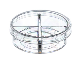
CELLview邃「 - Cell Culture Dish with Glass Bottom
Free of detectable DNase, RNase, human DNA
Non-pyrogenic, non-cytotoxic
Glass bottom features:
- High transparent achromatic
borosilicate glass; hydrolytic
class 1 (DIN ISO 719)
- Glass thickness 175 µm +/- 15 µm
- Maximal spectral transmission; no
autofluorescence
Advantages:
-Subdivided version enables
simultaneous multiplex analysis
- Embedded glass bottom for maximal
planarity
Number of compartments: 4
Diameter: 35 mm; height: 10 mm
Growth area: 1.9 cm²/compartment
Total volume: 1.5 ml/compartment
Working volume: 0.1 ml for seeding or staining only on glass area; 0.5 ml for cultivation in the complete compartment
Surface treatment: Advanced TC 邃「
Sterile
Quantity per bag/case: 10/40
Videos
Drug treatment during live cell imaging
A multi-position time-lapse experiment was started and after acquiring six time points every two minutes drugs were added to the different wells as indicated:
Video 1 - control (no drugs added)
In steady-state the Golgi apparatus is relatively stable on light microscopy level. The shape changes only slowly during the time of the experiment when observing control cells. Also the number of Golgi fragments visible by light microscopy resolution is relatively constant over time.Video 2 - Nocodazole added, final concentration 10 µM
Nocodazole treatment induces, fragmentation of the Golgi apparatus. The onset of fragmentation starts 10 to 15 minutes after addition of the drug. The onset of fragmentation differs between individual cells. Fragmentation of the central Golgi to many distributed ministacks is the final phenotype of microtubule depolymerization after three hours.Video 3 - Latrunculin B added, final concentration 1 ホシM
Actin depolymerization by Latrunculin B influences the shape of the Golgi from relatively thin elongated to a rounded up and compact appearance. After 10 to 20 minutes differences in the Golgi morphology became first visible and after approximately one hour the Golgi
rearrangement was completed.Video 4 - Brefeldin A added, final concentration 5 ホシg/ml
Block of export from the endoplasmatic reticulum (ER) by Brefeldin A leads to a rapid redistribution of the Golgi compartment to the ER by retrograde transport. This effect is often completed within 5 minutes.
Performing these experiments in parallel in CELLview邃「 dishes with four compartments it is possible to directly compare the speed and timing of drug effects on the Golgi apparatus. Brefeldin A affects Golgi morphology much faster than Nocodazole and Latrunculin B, which both induces first changes in the range of 10-20 minutes.


 粤公网安备44196802000105号